> Antigen, Antibodies, ELISA, Western Blot > Primary Antibody > Polyclonal Antibodies > Neurokinin Receptor 1 (NK1) (extracellular) AntibodyBrand |
Leading Biology | Catalog Number |
AMM06658G |
Product Type |
Polyclonal Antibodies | Field of Research |
|
Product Overview |
We constantly strive to ensure we provide our customers with the best antibodies. As a result of this work we offer this antibody in purified format.
We are in the process of updating our datasheets. If you have any questions regarding this update, please feel free to contact our technical support team.
This product is a high quality Neurokinin Receptor 1 (NK1) (extracellular) antibody.
|
||
Molecular Weight |
46366 Da
|
||
Host |
Rabbit
|
||
Species Reactivity |
Human, Mouse, Rat
|
||
Target |
Peptide CMIEWPEHPNRTYEK, corresponding to amino acid residues 180-194 of rat Neurokinin Receptor 1 (NK1) (Accession P14600). 2nd extracellular loop.
|
||
GeneID |
|||
UniProt ID |
|||
Summary |
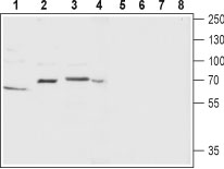
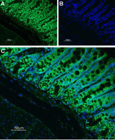
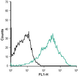
Substance P (SP), Neurokinin A (NKA) and Neurokinin B (NKB) are all peptides belonging to the Tachykinin protein family. These three peptides which demonstrate a quite heterogeneity in their distribution exert their effect via three receptors: Neurokinin 1-3 receptors, members of the G-protein coupled receptor superfamily. However, Neurokinin 1 Receptor (NK1) preferentially binds Substance P, Neurokinin 2 Receptor (NK2) to NKA and Neurokinin 3 Receptor (NK3) to NKB1.Neurokinin receptors are distinguished by their seven transmembrane domains, an extracellular N-terminus and a cytosolic C-terminal. An unusual property of these receptors is the presence of introns as part of their structural organization1-3. Tachykinin receptors undergo alternative splicing. For example, NK1 is detected with different C-terminal lengths. The longer receptor isoform is found in the brain whereas the truncated form is mostly detected in the periphery1,4.Due to the broad expression profile of tachykinin peptides, their respective receptors are also expressed in a similar fashion. NK1 is widely expressed in neurons endothelial cells, muscle and immune system cells. NK2 is broadly expressed in the periphery and its expression in the brain is quite restricted. NK3 on the other hand is largely expressed in the central nervous system and is also detected in the uterus, skeletal muscle, lung and liver1.Neurokinin receptors have been found in many pathophysiological indications and have therefore become targets for the development of pharmacological compounds. Such indications include cancer, psychological disorders, migraine and various inflammations, just to name a few5-8.Abgent is pleased to offer a highly specific antibody directed against an extracellular epitope of rat Neurokinin Receptor 1. Anti-Neurokinin Receptor 1 (NK1) (extracellular) antibody (#AG1005) can be used in western blot analysis and indirect flow cytometry applications, and has been designed to recognize NK1 from rat, mouse and human samples.
|
||
Form |
Affinity purified antibody, Liquid |
||
Storage & Stability |
Store at +4°C short term. For long-term storage, aliquot and store at -20°C or below. Stable for 12 months at -20°C. Avoid repeated freeze-thaw cycles.
|
||
Applications |
WB
|
||
Dilution |
WB~~1:200-1:2000
IHC~~1:100
|
||
Synonyms |
Substance-P receptor, SPR, NK-1 receptor, NK-1R, Tachykinin receptor 1, Tacr1, Tac1r
|
||
Images |
|
||
Specification |
|||
Quantity |
|
||
| Select | Brand | Catalog No. | Product Name | Pack Size | Type | Field of Research | Specification | Quantity | Price(USD) | |
| 1 | Leading Biology | APR03440G | ITGA11 Antibody (N-term) | 100 μl | Polyclonal Antibodies |
|
$495.00 | Add Ask | ||
| 2 | Leading Biology | APR04537G | CMIP Antibody (C-term) | 100 μl | Polyclonal Antibodies |
|
$495.00 | Add Ask | ||
| 3 | Leading Biology | APR12422G | Human H4 Histamine Receptor (extracellular) Antibody | 50 μl | Polyclonal Antibodies |
|
$695.00 | Add Ask | ||
| 4 | Leading Biology | APR03844G | UBE2W Antibody (C-term) | 100 μl | Polyclonal Antibodies |
|
$495.00 | Add Ask | ||
| 5 | Leading Biology | APR04349G | HECTD2 Antibody (N-term) | 100 μl | Polyclonal Antibodies |
|
$495.00 | Add Ask | ||
| 6 | Leading Biology | APR03502G | IGHG1 Antibody (Center) | 100 μl | Polyclonal Antibodies |
|
$495.00 | Add Ask |

You have 0 item in your cart
You have 0 item in your inquiry list