> Antigen, Antibodies, ELISA, Western Blot > Primary Antibody > Polyclonal Antibodies > MICA Antibody (Center)Brand |
Leading Biology | Catalog Number |
APR06106G |
Product Type |
Polyclonal Antibodies | Field of Research |
|
Product Overview |
We constantly strive to ensure we provide our customers with the best antibodies. As a result of this work we offer this antibody in purified format.
We are in the process of updating our datasheets. If you have any questions regarding this update, please feel free to contact our technical support team.
This product is a high quality MICA Antibody (Center).
|
||
Molecular Weight |
42915 Da
|
||
Cellular Localization |
Antigen Cellular Localization:
Cell membrane; Single- pass type I membrane protein Cytoplasm Note=Expressed on the cell surface in gastric epithelium, endothelial cells and fibroblasts and in the cytoplasm in keratinocytes and monocytes. Infection with human adenovirus 5 suppresses cell surface expression due to the adenoviral E3-19K protein which causes retention in the endoplasmic reticulum
|
||
Host |
Rabbit
|
||
Species Reactivity |
Human
|
||
Immunogen |
68-97 aa
|
||
Target |
This MICA antibody is generated from rabbits immunized with a KLH conjugated synthetic peptide between 68-97 amino acids from the Central region of human MICA.
|
||
Isotype |
Rabbit Ig
|
||
GeneID |
|||
UniProt ID |
|||
Function |
Seems to have no role in antigen presentation. Acts as a stress-induced self-antigen that is recognized by gamma delta T- cells. Ligand for the KLRK1/NKG2D receptor. Binding to KLRK1 leads to cell lysis.
|
||
Summary |
MICA is the higly polymorphic MHC (HLA) class I chain-related gene A. The protein product is expressed on the cell surface, although unlike canonical class I molecules does not seem to associate with beta-2-microglobulin. It is thought that MICA functions as a stress-induced antigen that is broadly recognized by intestinal epithelial gamma delta T cells.
|
||
Storage & Stability |
Store at +4°C short term. For long-term storage, aliquot and store at -20°C or below. Stable for 12 months at -20°C. Avoid repeated freeze-thaw cycles.
|
||
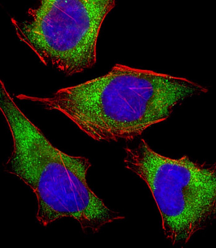
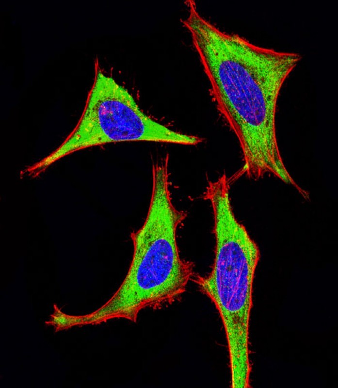
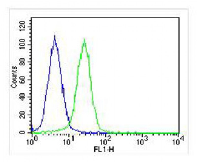
Applications |
WB, IHC-P, IF, FC, E
|
||
Dilution |
IF~~1:25
FC~~1:25
IHC-P~~1:25
WB~~1:1000
|
||
Images |
|
||
Specification |
|||
Quantity |
|
||
| Select | Brand | Catalog No. | Product Name | Pack Size | Type | Field of Research | Specification | Quantity | Price(USD) | |
| 1 | Leading Biology | APR03440G | ITGA11 Antibody (N-term) | 100 μl | Polyclonal Antibodies |
|
$495.00 | Add Ask | ||
| 2 | Leading Biology | APR04537G | CMIP Antibody (C-term) | 100 μl | Polyclonal Antibodies |
|
$495.00 | Add Ask | ||
| 3 | Leading Biology | APR12422G | Human H4 Histamine Receptor (extracellular) Antibody | 50 μl | Polyclonal Antibodies |
|
$695.00 | Add Ask | ||
| 4 | Leading Biology | APR03844G | UBE2W Antibody (C-term) | 100 μl | Polyclonal Antibodies |
|
$495.00 | Add Ask | ||
| 5 | Leading Biology | APR04349G | HECTD2 Antibody (N-term) | 100 μl | Polyclonal Antibodies |
|
$495.00 | Add Ask | ||
| 6 | Leading Biology | APR03502G | IGHG1 Antibody (Center) | 100 μl | Polyclonal Antibodies |
|
$495.00 | Add Ask |

You have 0 item in your cart
You have 0 item in your inquiry list