> Antigen, Antibodies, ELISA, Western Blot > Primary Antibody > Polyclonal Antibodies > Ryanodine Receptor 2 AntibodyBrand |
Leading Biology | Catalog Number |
AMR09796G |
Product Type |
Polyclonal Antibodies | Field of Research |
|
Product Overview |
We constantly strive to ensure we provide our customers with the best antibodies. As a result of this work we offer this antibody in purified format.
We are in the process of updating our datasheets. If you have any questions regarding this update, please feel free to contact our technical support team.
This product is a high quality Ryanodine Receptor 2 antibody.
|
||
Molecular Weight |
564567 Da
|
||
Host |
Rabbit
|
||
Species Reactivity |
Mouse, Rat
|
||
Target |
Peptide CAGESMSPGQGRNN, corresponding to amino acid residues 1489-1502 of human Ryanodine Receptor 2 (Accession Q92736). Intracellular, N-terminus.
|
||
GeneID |
|||
UniProt ID |
|||
Summary |
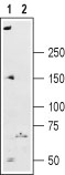
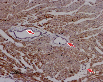
It is well established that cytosolic calcium (Ca2+) acts as a key second messenger in many intracellular pathways including synaptic transmission, muscle contraction, hormonal secretion, cell growth and proliferation.1,2 The primary intracellular Ca2+ storage/release organelle in most cells is the endoplasmic reticulum (ER) or the sarcoplasmic reticulum (SR) in striated muscle cells.The ER and SR contain two Ca2+ release channels families, the Inositol trisphosphate receptors (IP3Rs) and the Ryanodine receptors (RyRs).3The Ryanodine receptor family consists of three different isoforms: The skeletal muscleisoform, Ryanodine Receptor type 1 (RyR1); the cardiac muscle isoform, Ryanodine Receptor type 2 (RyR2) and the brain isoform, Ryanodine Receptor type 3 (RyR3).3 The Ryanodine receptors are homotetrameric proteins. They play a key role in the mechanism of excitation-contraction coupling in striated muscle. Binding of Ryanodine to the Ryanodine Receptor causes to two major changes in the channel: a reduction in single-channel conductance and a marked increase in open state probability.RyR2 serves as an intracellular Ca2+ channel in the SR membrane. It is predominantly expressed in cardiac muscle where it plays a central role in cardiac excitation-contraction coupling. RyR2 is also expressed in the brain. 1-4Abgent is pleased to offer a highly specific antibody directed against an epitope located at thecytoplasmicregion of the human Ryanodine receptor 2 (RyR2). The Anti-Ryanodine Receptor 2 antibody (#AG1394) can be used in western blot analysis and Immunohistochemical applications. It has been designed to recognize RyR2 from human, rat and mouse samples.
|
||
Form |
Affinity purified antibody, Liquid |
||
Storage & Stability |
Store at +4°C short term. For long-term storage, aliquot and store at -20°C or below. Stable for 12 months at -20°C. Avoid repeated freeze-thaw cycles.
|
||
Applications |
WB
|
||
Dilution |
WB~~1:200-1:2000
IHC~~1:100
|
||
Synonyms |
Ryanodine receptor 2, RYR-2, RyR2, hRYR-2, Cardiac muscle ryanodine receptor, Cardiac muscle ryanodine receptor-calcium release channel, Type 2 ryanodine receptor, RYR2
|
||
Images |
|
||
Specification |
|||
Quantity |
|
||
| Select | Brand | Catalog No. | Product Name | Pack Size | Type | Field of Research | Specification | Quantity | Price(USD) | |
| 1 | Leading Biology | APR03440G | ITGA11 Antibody (N-term) | 100 μl | Polyclonal Antibodies |
|
$495.00 | Add Ask | ||
| 2 | Leading Biology | APR04537G | CMIP Antibody (C-term) | 100 μl | Polyclonal Antibodies |
|
$495.00 | Add Ask | ||
| 3 | Leading Biology | APR12422G | Human H4 Histamine Receptor (extracellular) Antibody | 50 μl | Polyclonal Antibodies |
|
$695.00 | Add Ask | ||
| 4 | Leading Biology | APR03844G | UBE2W Antibody (C-term) | 100 μl | Polyclonal Antibodies |
|
$495.00 | Add Ask | ||
| 5 | Leading Biology | APR04349G | HECTD2 Antibody (N-term) | 100 μl | Polyclonal Antibodies |
|
$495.00 | Add Ask | ||
| 6 | Leading Biology | APR03502G | IGHG1 Antibody (Center) | 100 μl | Polyclonal Antibodies |
|
$495.00 | Add Ask |

You have 0 item in your cart
You have 0 item in your inquiry list