> Antigen, Antibodies, ELISA, Western Blot > Primary Antibody > Polyclonal Antibodies > ATG7 Antibody (C-term)Brand |
Leading Biology | Catalog Number |
APR10816G |
Product Type |
Polyclonal Antibodies | Field of Research |
|
Product Overview |
We constantly strive to ensure we provide our customers with the best antibodies. As a result of this work we offer this antibody in purified format.
We are in the process of updating our datasheets. If you have any questions regarding this update, please feel free to contact our technical support team.
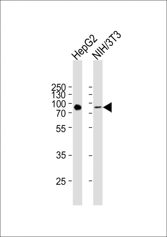
This product is a high quality ATG7 antibody (C-term).
|
||
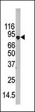
Molecular Weight |
77960 Da
|
||
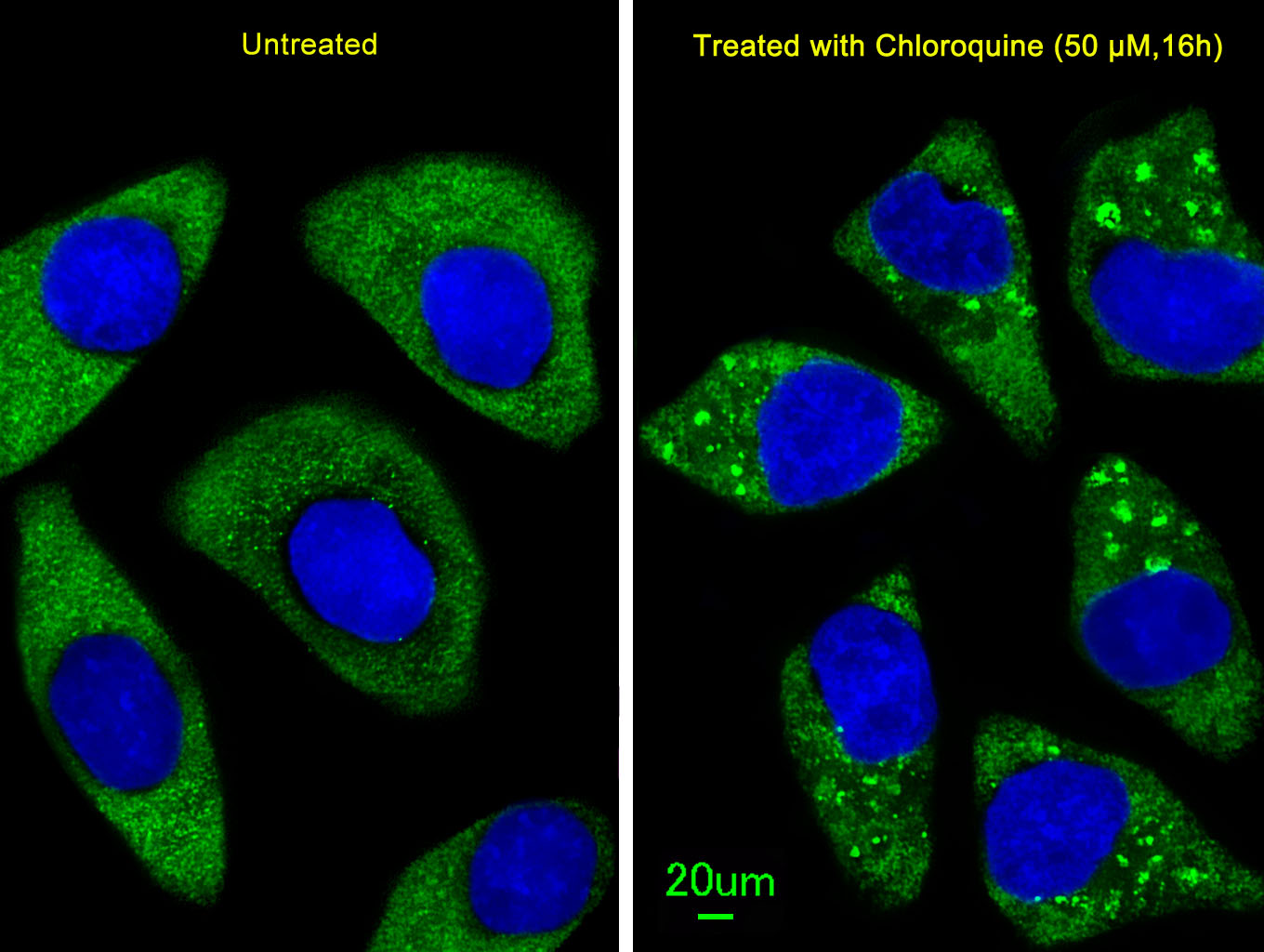
Cellular Localization |
Antigen Cellular Localization:
Cytoplasm. Preautophagosomal structure. Note=Localizes also to discrete punctae along the ciliary axoneme and to the base of the ciliary axoneme
|
||
Host |
Rabbit
|
||
Species Reactivity |
Human, Mouse
|
||
Immunogen |
540-569 aa
|
||
Target |
This ATG7 antibody is generated from rabbits immunized with a KLH conjugated synthetic peptide between 540-569 amino acids from the C-terminal region of human ATG7.
|
||
Isotype |
Rabbit Ig
|
||
Symbol |
APG7L
|
||
GeneID |
|||
UniProt ID |
|||
Function |
E1-like activating enzyme involved in the 2 ubiquitin- like systems required for cytoplasm to vacuole transport (Cvt) and autophagy. Activates ATG12 for its conjugation with ATG5 as well as the ATG8 family proteins for their conjugation with phosphatidylethanolamine. Both systems are needed for the ATG8 association to Cvt vesicles and autophagosomes membranes. Required for autophagic death induced by caspase-8 inhibition. Required for mitophagy which contributes to regulate mitochondrial quantity and quality by eliminating the mitochondria to a basal level to fulfill cellular energy requirements and preventing excess ROS production. Modulates p53/TP53 activity to regulate cell cycle and survival during metabolic stress. Plays also a key role in the maintenance of axonal homeostasis, the prevention of axonal degeneration, the maintenance of hematopoietic stem cells, the formation of Paneth cell granules, as well as in adipose differentiation.
|
||
Summary |
Macroautophagy is the major inducible pathway for the general turnover of cytoplasmic constituents in eukaryotic cells, it is also responsible for the degradation of active cytoplasmic enzymes and organelles during nutrient starvation. Macroautophagy involves the formation of double-membrane bound autophagosomes which enclose the cytoplasmic constituent targeted for degradation in a membrane bound structure, which then fuse with the lysosome (or vacuole) releasing a single-membrane bound autophagic bodies which are then degraded within the lysosome (or vacuole). APG7 functions as an E1 enzyme essential for multisubstrates such as GABARAPL1 and ATG12. APG3L is an E2-like conjugating enzyme facilitating covalent binding of APG8 (MAP1LC3) to phosphatidylethanolamine (PE). APG7 (an E1-like enzyme) facilitates this reaction by forming an E1-E2 complex with APG3. Formation of the PE conjugate is essential for autophagy.
|
||
Form |
Purified polyclonal antibody supplied in PBS with 0.09% (W/V) sodium azide. This antibody is purified through a protein A column, followed by peptide affinity purification. |
||
Storage & Stability |
Store at +4°C short term. For long-term storage, aliquot and store at -20°C or below. Stable for 12 months at -20°C. Avoid repeated freeze-thaw cycles.
|
||
Applications |
WB, IHC-P, IF, E
|
||
Dilution |
WB~~1:1000
IF~~1:25
IHC-P~~1:50~100
|
||
Synonyms |
Ubiquitin-like modifier-activating enzyme ATG7, ATG12-activating enzyme E1 ATG7, Autophagy-related protein 7, APG7-like, hAGP7, Ubiquitin-activating enzyme E1-like protein, ATG7, APG7L
|
||
Images |
|
||
Specification |
|||
Quantity |
|
||
| Select | Brand | Catalog No. | Product Name | Pack Size | Type | Field of Research | Specification | Quantity | Price(USD) | |
| 1 | Leading Biology | APR03440G | ITGA11 Antibody (N-term) | 100 μl | Polyclonal Antibodies |
|
$495.00 | Add Ask | ||
| 2 | Leading Biology | APR04537G | CMIP Antibody (C-term) | 100 μl | Polyclonal Antibodies |
|
$495.00 | Add Ask | ||
| 3 | Leading Biology | APR12422G | Human H4 Histamine Receptor (extracellular) Antibody | 50 μl | Polyclonal Antibodies |
|
$695.00 | Add Ask | ||
| 4 | Leading Biology | APR03844G | UBE2W Antibody (C-term) | 100 μl | Polyclonal Antibodies |
|
$495.00 | Add Ask | ||
| 5 | Leading Biology | APR04349G | HECTD2 Antibody (N-term) | 100 μl | Polyclonal Antibodies |
|
$495.00 | Add Ask | ||
| 6 | Leading Biology | APR03502G | IGHG1 Antibody (Center) | 100 μl | Polyclonal Antibodies |
|
$495.00 | Add Ask |

You have 0 item in your cart
You have 0 item in your inquiry list