> Antigen, Antibodies, ELISA, Western Blot > Primary Antibody > Polyclonal Antibodies > EphA1 (extracellular) AntibodyBrand |
Leading Biology | Catalog Number |
APR11842G |
Product Type |
Polyclonal Antibodies | Field of Research |
|
Product Overview |
We constantly strive to ensure we provide our customers with the best antibodies. As a result of this work we offer this antibody in purified format.
We are in the process of updating our datasheets. If you have any questions regarding this update, please feel free to contact our technical support team.
This product is a high quality EphA1 (extracellular) antibody.
|
||
Molecular Weight |
108127 Da
|
||
Host |
Rabbit
|
||
Species Reactivity |
Human, Mouse, Rat
|
||
Target |
Peptide (C)KKEPRQLELTWAGSR, corresponding to amino acid residues 457-471 of human EphA1 (Accession P21709). Extracellular, N-terminus.
|
||
GeneID |
|||
UniProt ID |
|||
Summary |
Eph receptors are the largest family of receptor tyrosine kinases (RTKs). EphA receptors (EphA1-10) bind ephrinA ligands which are GPI-linked proteins and EphB receptors (EphB1-6) bind ephrinB ligands which are membrane protein with one transmembrane domain1,2. Within each subfamily, interactions between receptor and ligand are promiscuous. In addition, Eph receptors can also bind ephrins from the other class2. Forward and reverse signaling through Eph receptors is a unique characteristic to this RTK since ephrins are physically linked to the plasma membrane3.Structurally, Eph receptors contain an extracellular ligand-binding domain, a transmembrane domain and an intracellular C-terminal domain responsible for intracellular signaling1. Forward Eph receptor signaling involves autophosphorylation of the receptor via a tyrosine kinase domain, as well as phosphorylation of other proteins. Known effectors of the forward signaling include Src kinase and Ras/Rho GTPases2. Much less is known about the reverse signaling mediated by Eph receptors. Besides from acting independently, Eph receptors can also signal in concert with other receptors. For example, Eph receptors cooperate with FGF receptor, NMDA ligand-gated ion channel and chemokine G-protein coupled receptor2.Biological activities attributed to the Eph receptor-ephrin signaling module include establishing neuronal connections, mediating neuronal plasticity and repair following neuronal injury2,4. Eph receptors may also have a role in the immune system5.Eph receptors are expressed in the developing nervous system, and in the adult brain. It is also detected in the pancreas, intestine, bone and lymphocytes. In cancer cells, Eph receptors and ephrins are overexpressed2,6,7. They are also implicated in neurodegenerative disorders like Alzheimer’s disease1.Abgent is pleased to offer a highly specific antibody directed against an extracellular epitope of human EphA1. Anti-EphA1 (extracellular) antibody (#AG1283) can be used in western blot, immunocytochemistry and indirect flow cytometry applications. It has been designed to recognize EphA1 from rat, mouse, and human samples.
|
||
Form |
Affinity purified antibody, Liquid |
||
Storage & Stability |
Store at +4°C short term. For long-term storage, aliquot and store at -20°C or below. Stable for 12 months at -20°C. Avoid repeated freeze-thaw cycles.
|
||
Applications |
WB, LCI
|
||
Dilution |
WB~~1:200-1:2000
ICC~~1:25
FC~~1:20
|
||
Synonyms |
Ephrin type-A receptor 1, hEpha1, EPH tyrosine kinase, EPH tyrosine kinase 1, Erythropoietin-producing hepatoma receptor, Tyrosine-protein kinase receptor EPH, EPHA1, EPH, EPHT, EPHT1
|
||
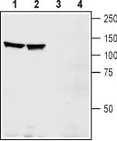
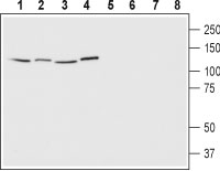
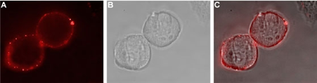
Images |
|
||
Specification |
|||
Quantity |
|
||
| Select | Brand | Catalog No. | Product Name | Pack Size | Type | Field of Research | Specification | Quantity | Price(USD) | |
| 1 | Leading Biology | APR03440G | ITGA11 Antibody (N-term) | 100 μl | Polyclonal Antibodies |
|
$495.00 | Add Ask | ||
| 2 | Leading Biology | APR04537G | CMIP Antibody (C-term) | 100 μl | Polyclonal Antibodies |
|
$495.00 | Add Ask | ||
| 3 | Leading Biology | APR12422G | Human H4 Histamine Receptor (extracellular) Antibody | 50 μl | Polyclonal Antibodies |
|
$695.00 | Add Ask | ||
| 4 | Leading Biology | APR03844G | UBE2W Antibody (C-term) | 100 μl | Polyclonal Antibodies |
|
$495.00 | Add Ask | ||
| 5 | Leading Biology | APR04349G | HECTD2 Antibody (N-term) | 100 μl | Polyclonal Antibodies |
|
$495.00 | Add Ask | ||
| 6 | Leading Biology | APR03502G | IGHG1 Antibody (Center) | 100 μl | Polyclonal Antibodies |
|
$495.00 | Add Ask |

You have 0 item in your cart
You have 0 item in your inquiry list