> Antigen, Antibodies, ELISA, Western Blot > Primary Antibody > Polyclonal Antibodies > D3 Dopamine Receptor (extracellular) AntibodyBrand |
Leading Biology | Catalog Number |
APG03236G |
Product Type |
Polyclonal Antibodies | Field of Research |
|
Product Overview |
We constantly strive to ensure we provide our customers with the best antibodies. As a result of this work we offer this antibody in purified format.
We are in the process of updating our datasheets. If you have any questions regarding this update, please feel free to contact our technical support team.
This product is a high quality D3 Dopamine Receptor (extracellular) antibody.
|
||
Molecular Weight |
49516 Da
|
||
Host |
Rabbit
|
||
Species Reactivity |
Mouse, Rat
|
||
Target |
Peptide CGAENSTGVNRARPH, corresponding to amino acid residues 15-29 of rat D3 Dopamine receptor (Accession P19020). Extracellular, N-terminus.
|
||
GeneID |
|||
UniProt ID |
|||
Summary |
The D3 Dopamine Receptor (D3 receptor) is one of five receptors that mediate the effects of the catecholamine neurotransmitter dopamine. Dopamine regulates a variety of functions including locomotor activity, emotion, positive reinforcement, food intake, and endocrine regulation. The dopaminergic system has been extensively studied in the last thirty years mainly because its dysregulation has been linked to several neurological and neuropsychiatric diseases including Parkinson’s disease and schizophrenia.1 All five dopamine receptors belong to the 7-transmembrane domain, G protein-coupled receptor (GPCR) superfamily. Historically, the five receptors have been divided into two subfamilies based on pharmacological and structural considerations: the D1-like subfamily (that includes the D1 and D5 subtypes) and the D2-like subfamily (that includes the D2-, D3- and D4 subtypes).1The D1-like receptors are coupled to Gs-type G proteins and enhance adenylate cyclase activity while the D2-like receptors are coupled to Gi-type G proteins and inhibit adenylate cyclase activity.1 The D3 receptor distribution in the brain is relatively restricted to limbic areas such as striatum, islands of Calleja and olfactory tubercle. In the periphery, it is expressed in the kidney particularly in proximal tubules.1 The exact physiological function of the D3 receptor remains to be fully elucidated. In studies using D3 receptor knockout mice, the most prominent dysfunction was the development of rennin-dependent hypertension. Potential roles in reinforcement and reward behaviors have also been suggested as well as roles in neuropsychiatric disorders such as drug abuse and schizophrenia.2 Abgent is pleased to offer a highly specific antibody directed against an epitope located at the extracellular N-terminus domain of the rat D3 dopamine receptor. Anti-D3 Dopamine Receptor (extracellular) antibody (#AG1289) can be used in western blot analysis and immunohistochemical applications, and recognizes D3 dopamine receptor from rat and mouse samples
|
||
Form |
Affinity purified antibody, Liquid |
||
Storage & Stability |
Store at +4°C short term. For long-term storage, aliquot and store at -20°C or below. Stable for 12 months at -20°C. Avoid repeated freeze-thaw cycles.
|
||
Applications |
WB, IHC
|
||
Dilution |
WB~~1:200-1:2000
IHC~~1:100
|
||
Synonyms |
D(3) dopamine receptor, Dopamine D3 receptor, Drd3
|
||
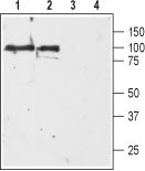
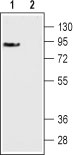
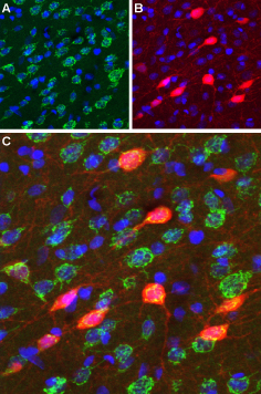
Images |
|
||
Specification |
|||
Quantity |
|
||
| Select | Brand | Catalog No. | Product Name | Pack Size | Type | Field of Research | Specification | Quantity | Price(USD) | |
| 1 | Leading Biology | APR03440G | ITGA11 Antibody (N-term) | 100 μl | Polyclonal Antibodies |
|
$495.00 | Add Ask | ||
| 2 | Leading Biology | APR04537G | CMIP Antibody (C-term) | 100 μl | Polyclonal Antibodies |
|
$495.00 | Add Ask | ||
| 3 | Leading Biology | APR12422G | Human H4 Histamine Receptor (extracellular) Antibody | 50 μl | Polyclonal Antibodies |
|
$695.00 | Add Ask | ||
| 4 | Leading Biology | APR03844G | UBE2W Antibody (C-term) | 100 μl | Polyclonal Antibodies |
|
$495.00 | Add Ask | ||
| 5 | Leading Biology | APR04349G | HECTD2 Antibody (N-term) | 100 μl | Polyclonal Antibodies |
|
$495.00 | Add Ask | ||
| 6 | Leading Biology | APR03502G | IGHG1 Antibody (Center) | 100 μl | Polyclonal Antibodies |
|
$495.00 | Add Ask |

You have 0 item in your cart
You have 0 item in your inquiry list