> Antigen, Antibodies, ELISA, Western Blot > Primary Antibody > Polyclonal Antibodies > GABA (A) alpha1 Receptor (extracellular) AntibodyBrand |
Leading Biology | Catalog Number |
APR11953G |
Product Type |
Polyclonal Antibodies | Field of Research |
|
Product Overview |
We constantly strive to ensure we provide our customers with the best antibodies. As a result of this work we offer this antibody in purified format.
We are in the process of updating our datasheets. If you have any questions regarding this update, please feel free to contact our technical support team.
This product is a high quality GABA (A) alpha1 Receptor (extracellular) antibody.
|
||
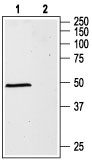
Molecular Weight |
51754 Da
|
||
Host |
Rabbit
|
||
Species Reactivity |
Mouse, Rat
|
||
Target |
Peptide QPSQDELKDNTTVFTR(C), corresponding to amino acid residues 28-43 of rat GABA (A) ?±1 Receptor (Accession P62813). Extracellular, N-terminus.
|
||
GeneID |
|||
UniProt ID |
|||
Summary |
GABA (γ-aminobutyric acid) is the major inhibitory neurotransmitter in the brain. Its production, release, reuptake, and metabolism all occur in the nervous system.1The GABA transmitter interacts with two major types of receptors: ionotropic GABAA receptors (GABAAR) and metabotropic receptors (GABABR). GABAARs belong to the ligand-gated ion channel superfamily.2 GABA inhibits the activity of signal-receiving neurons by interacting with the GABAA receptor on these cells.3 Binding of GABA to its GABAA receptor results in conformational changes that open a Cl- channel, producing an increase in membrane conductance that results in inhibition of neural activity.2GABAARs are heteropentamers, in which all five subunits contribute to pore formation. To date, eight subunit isoforms have been cloned:α, β, γ, δ, ε, π, θ, and ρ.1 Six α subunit isoforms have been found to exist in mammals (α1-α6). In most cases, native GABAA receptors consists of 2α, 2β, and 1δ subunits. The α subunit is the most common and is expressed ubiquitously. It determines the affinities of GABAARs for allosteric ligands.Each subtype has a unique regional expression in the brain, and individual neurons often express multiple subtypes.4 The α1 subunit is highly expressed in adulthood while the α2 subunit is highly expressed very early in rat brain development. Failure to complete the normal transition between the α-subunits that are highly expressed in early development (α2, α3, and α5) and those expressed in adulthood (α1) is suggested to play a major role in the development of temporal lobe epilepsy.5
|
||
Form |
Affinity purified antibody, Liquid |
||
Storage & Stability |
Store at +4°C short term. For long-term storage, aliquot and store at -20°C or below. Stable for 12 months at -20°C. Avoid repeated freeze-thaw cycles.
|
||
Applications |
WB, LCI
|
||
Dilution |
WB~~1:200-1:2000
|
||
Synonyms |
Gamma-aminobutyric acid receptor subunit alpha-1, GABA(A) receptor subunit alpha-1, Gabra1, Gabra-1
|
||
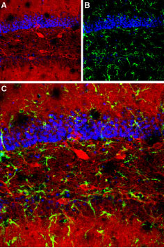
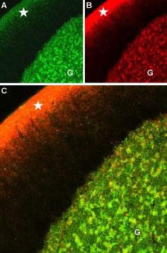
Images |
|
||
Specification |
|||
Quantity |
|
||
| Select | Brand | Catalog No. | Product Name | Pack Size | Type | Field of Research | Specification | Quantity | Price(USD) | |
| 1 | Leading Biology | APR03440G | ITGA11 Antibody (N-term) | 100 μl | Polyclonal Antibodies |
|
$495.00 | Add Ask | ||
| 2 | Leading Biology | APR04537G | CMIP Antibody (C-term) | 100 μl | Polyclonal Antibodies |
|
$495.00 | Add Ask | ||
| 3 | Leading Biology | APR12422G | Human H4 Histamine Receptor (extracellular) Antibody | 50 μl | Polyclonal Antibodies |
|
$695.00 | Add Ask | ||
| 4 | Leading Biology | APR03844G | UBE2W Antibody (C-term) | 100 μl | Polyclonal Antibodies |
|
$495.00 | Add Ask | ||
| 5 | Leading Biology | APR04349G | HECTD2 Antibody (N-term) | 100 μl | Polyclonal Antibodies |
|
$495.00 | Add Ask | ||
| 6 | Leading Biology | APR03502G | IGHG1 Antibody (Center) | 100 μl | Polyclonal Antibodies |
|
$495.00 | Add Ask |

You have 0 item in your cart
You have 0 item in your inquiry list