> Antigen, Antibodies, ELISA, Western Blot > Primary Antibody > Polyclonal Antibodies > K2P18.1 (TRESK) (extracellular) AntibodyBrand |
Leading Biology | Catalog Number |
AMM06052G |
Product Type |
Polyclonal Antibodies | Field of Research |
|
Product Overview |
We constantly strive to ensure we provide our customers with the best antibodies. As a result of this work we offer this antibody in purified format.
We are in the process of updating our datasheets. If you have any questions regarding this update, please feel free to contact our technical support team.
This product is a high quality K2P18.1 (TRESK) (extracellular) antibody.
|
||
Molecular Weight |
44403 Da
|
||
Host |
Rabbit
|
||
Species Reactivity |
Mouse, Rat
|
||
Target |
Peptide (C)EAEENPELKKFLDD, corresponding to amino acid residues 62-75 of mouse K2P18.1 (Accession Q6VV64 ).??1st extracellular loop.Unlikely to recognize human samples.
|
||
GeneID |
|||
UniProt ID |
|||
Summary |
K2P18.1 (also named TWIK-related spinal cord K+ channel, TRESK or KCNK18) is a member of the 2-pore (2P) domain K+ channels family that in mammals includes 15 members. These channels show little time or voltage dependence and are considered to be “leak” or “background” K+ channels, thereby generating background currents which help set the membrane resting potential and control cell excitation.1The K2P channels have a signature topology that includes four transmembrane domains and two pore domains with intracellular N- and C termini. It has been proposed that the functional channel unit is a dimer.Different K2Pfamily membersare regulated by diverse physical and chemical stimuli including temperature, pH, mechanical stretch, inhalation anesthetics, signaling pathways (PKC and PKA), arachidonic acid, etc.K2P18.1 is the only K2P channel so far, whose current is activated following Gaq-receptor coupled activation. The enhancement of K2P18.1 current involves activation of calcineurin (calcium–calmodulin-dependent phosphatase 2B) following the rise in intracellular calcium that occurs subsequent to Gaq activation.2 In addition, K2P18.1 is potently activated by clinical concentrations of volatile anesthetics.3K2P18.1 expression in humans is largely restricted to the spinal cord although in rodents it has a broader expression pattern that includes brain, testis and spleen.K2P18.1 represents the most important background K+ channel in dorsal root ganglion neurons and hence it has been postulated that it has an important role in acute and chronic pain as well as general anesthesia.Abgent is pleased to offer a highly specific Anti-K2P18.1 (TRESK) (extracellular) antibody (#AG1091) directed against a well conserved epitope located in the first extracellular region of the mouse K2P18.1 channel.Anti-K2P18.1 (TRESK) (extracellular) antibody can be used in western blot, immunohistochemical, and immunocytochemical applications and will recognize K2P18.1 from rat and mouse samples.
|
||
Form |
Affinity purified antibody, Liquid |
||
Storage & Stability |
Store at +4°C short term. For long-term storage, aliquot and store at -20°C or below. Stable for 12 months at -20°C. Avoid repeated freeze-thaw cycles.
|
||
Applications |
WB
|
||
Dilution |
WB~~1:200-1:2000
IHC~~1:100
ICC~~1:50
|
||
Synonyms |
Potassium channel subfamily K member 18, Two-pore-domain potassium channel TRESK, Kcnk18, Tresk-2, Tresk2
|
||
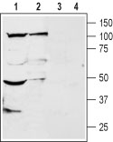
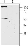
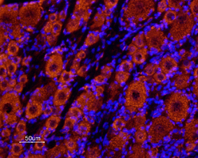
Images |
|
||
Specification |
|||
Quantity |
|
||
| Select | Brand | Catalog No. | Product Name | Pack Size | Type | Field of Research | Specification | Quantity | Price(USD) | |
| 1 | Leading Biology | APR03440G | ITGA11 Antibody (N-term) | 100 μl | Polyclonal Antibodies |
|
$495.00 | Add Ask | ||
| 2 | Leading Biology | APR04537G | CMIP Antibody (C-term) | 100 μl | Polyclonal Antibodies |
|
$495.00 | Add Ask | ||
| 3 | Leading Biology | APR12422G | Human H4 Histamine Receptor (extracellular) Antibody | 50 μl | Polyclonal Antibodies |
|
$695.00 | Add Ask | ||
| 4 | Leading Biology | APR03844G | UBE2W Antibody (C-term) | 100 μl | Polyclonal Antibodies |
|
$495.00 | Add Ask | ||
| 5 | Leading Biology | APR04349G | HECTD2 Antibody (N-term) | 100 μl | Polyclonal Antibodies |
|
$495.00 | Add Ask | ||
| 6 | Leading Biology | APR03502G | IGHG1 Antibody (Center) | 100 μl | Polyclonal Antibodies |
|
$495.00 | Add Ask |

You have 0 item in your cart
You have 0 item in your inquiry list