> Antigen, Antibodies, ELISA, Western Blot > Primary Antibody > Polyclonal Antibodies > p44/42 MAPK (Erk1/2) AntibodyBrand |
Leading Biology | Catalog Number |
APR12683G |
Product Type |
Polyclonal Antibodies | Field of Research |
|
Product Overview |
We constantly strive to ensure we provide our customers with the best antibodies. As a result of this work we offer this antibody in purified format.
We are in the process of updating our datasheets. If you have any questions regarding this update, please feel free to contact our technical support team.
This product is a high quality p44/42 MAPK (Erk1/2) antibody.
|
||
Molecular Weight |
41276 Da
|
||
Cellular Localization |
Antigen Cellular Localization:
Cytoplasm, cytoskeleton, spindle. Nucleus. Cytoplasm, cytoskeleton, microtubule organizing center, centrosome. Cytoplasm. Note=Associated with the spindle during prometaphase and metaphase. PEA15-binding and phosphorylated DAPK1 promote its cytoplasmic retention. Phosphorylation at Ser- 244 and Ser-246 as well as autophosphorylation at Thr-188 promote nuclear localization.
|
||
Host |
Rabbit
|
||
Species Reactivity |
Human, Mouse, Rat, Chicken, Sheep
|
||
Target |
p44/42 MAPK (Erk1/2)
|
||
Isotype |
Rabbit IgG
|
||
Symbol |
Erk2, Mapk, Prkm1
|
||
GeneID |
|||
UniProt ID |
|||
Function |
Serine/threonine kinase which acts as an essential component of the MAP kinase signal transduction pathway. MAPK1/ERK2 and MAPK3/ERK1 are the 2 MAPKs which play an important role in the MAPK/ERK cascade. They participate also in a signaling cascade initiated by activated KIT and KITLG/SCF. Depending on the cellular context, the MAPK/ERK cascade mediates diverse biological functions such as cell growth, adhesion, survival and differentiation through the regulation of transcription, translation, cytoskeletal rearrangements. The MAPK/ERK cascade plays also a role in initiation and regulation of meiosis, mitosis, and postmitotic functions in differentiated cells by phosphorylating a number of transcription factors. About 160 substrates have already been discovered for ERKs. Many of these substrates are localized in the nucleus, and seem to participate in the regulation of transcription upon stimulation. However, other substrates are found in the cytosol as well as in other cellular organelles, and those are responsible for processes such as translation, mitosis and apoptosis. Moreover, the MAPK/ERK cascade is also involved in the regulation of the endosomal dynamics, including lysosome processing and endosome cycling through the perinuclear recycling compartment (PNRC); as well as in the fragmentation of the Golgi apparatus during mitosis. The substrates include transcription factors (such as ATF2, BCL6, ELK1, ERF, FOS, HSF4 or SPZ1), cytoskeletal elements (such as CANX, CTTN, GJA1, MAP2, MAPT, PXN, SORBS3 or STMN1), regulators of apoptosis (such as BAD, BTG2, CASP9, DAPK1, IER3, MCL1 or PPARG), regulators of translation (such as EIF4EBP1) and a variety of other signaling-related molecules (like ARHGEF2, DCC, FRS2 or GRB10). Protein kinases (such as RAF1, RPS6KA1/RSK1, RPS6KA3/RSK2, RPS6KA2/RSK3, RPS6KA6/RSK4, SYK, MKNK1/MNK1, MKNK2/MNK2, RPS6KA5/MSK1, RPS6KA4/MSK2, MAPKAPK3 or MAPKAPK5) and phosphatases (such as DUSP1, DUSP4, DUSP6 or DUSP16) are other substrates which enable the propagation the MAPK/ERK signal to additional cytosolic and nuclear targets, thereby extending the specificity of the cascade. Mediates phosphorylation of TPR in respons to EGF stimulation. May play a role in the spindle assembly checkpoint. Phosphorylates PML and promotes its interaction with PIN1, leading to PML degradation (By similarity).
|
||
Summary |
The extracellular signal-regulated kinase 1 and 2 (Erk1 and Erk2) are closely related mitogen activated protein (MAP) kinases that are activated thro μgh the extracellular stimulation by many growth factors, mitogens and differentiation promoting agents via a protein kinase cascade. Erk1/2 are activated approximately 1000 fold by phosphorylation of neighboring threonine and tyrosine residues by Mek1 and Mek2. Both sites must be phosphorylated for maximum activity. Erk1/2 kinases are ubiquitously distributed in the nervous system, as well as a broad range of cells and tissues. These two kinases represent proximal kinases in the classical MAP kinase cascade pathway which links growth and differentiation signals at the cell surface (thro μgh tyrosine kinase) with transcription in the nucleus.
|
||
Form |
Liquid |
||
Storage & Stability |
Store at +4°C short term. For long-term storage, aliquot and store at -20°C or below. Stable for 12 months at -20°C. Avoid repeated freeze-thaw cycles.
|
||
Applications |
WB
|
||
Synonyms |
MAPK1, p38, ERK , P42MAPK, ERK2 , p40 , PRKM1 , p41mapk , PRKM2, MAPK2 , p42-MAPK, p41 , ERK-2, ERT1
|
||
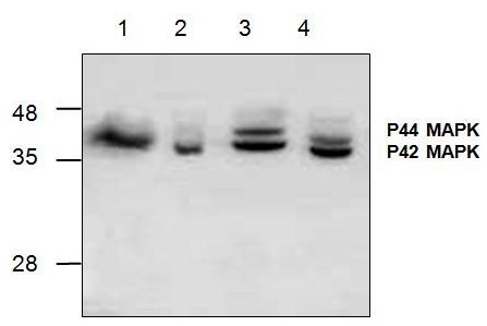
Images |
|
||
Specification |
|||
Quantity |
|
||
| Select | Brand | Catalog No. | Product Name | Pack Size | Type | Field of Research | Specification | Quantity | Price(USD) | |
| 1 | Leading Biology | APR03440G | ITGA11 Antibody (N-term) | 100 μl | Polyclonal Antibodies |
|
$495.00 | Add Ask | ||
| 2 | Leading Biology | APR04537G | CMIP Antibody (C-term) | 100 μl | Polyclonal Antibodies |
|
$495.00 | Add Ask | ||
| 3 | Leading Biology | APR12422G | Human H4 Histamine Receptor (extracellular) Antibody | 50 μl | Polyclonal Antibodies |
|
$695.00 | Add Ask | ||
| 4 | Leading Biology | APR03844G | UBE2W Antibody (C-term) | 100 μl | Polyclonal Antibodies |
|
$495.00 | Add Ask | ||
| 5 | Leading Biology | APR04349G | HECTD2 Antibody (N-term) | 100 μl | Polyclonal Antibodies |
|
$495.00 | Add Ask | ||
| 6 | Leading Biology | APR03502G | IGHG1 Antibody (Center) | 100 μl | Polyclonal Antibodies |
|
$495.00 | Add Ask |

You have 0 item in your cart
You have 0 item in your inquiry list