> Antigen, Antibodies, ELISA, Western Blot > Primary Antibody > Polyclonal Antibodies > SPHK1 Antibody (Center)Brand |
Leading Biology | Catalog Number |
APR06976G |
Product Type |
Polyclonal Antibodies | Field of Research |
|
Product Overview |
We constantly strive to ensure we provide our customers with the best antibodies. As a result of this work we offer this antibody in purified format.
We are in the process of updating our datasheets. If you have any questions regarding this update, please feel free to contact our technical support team.
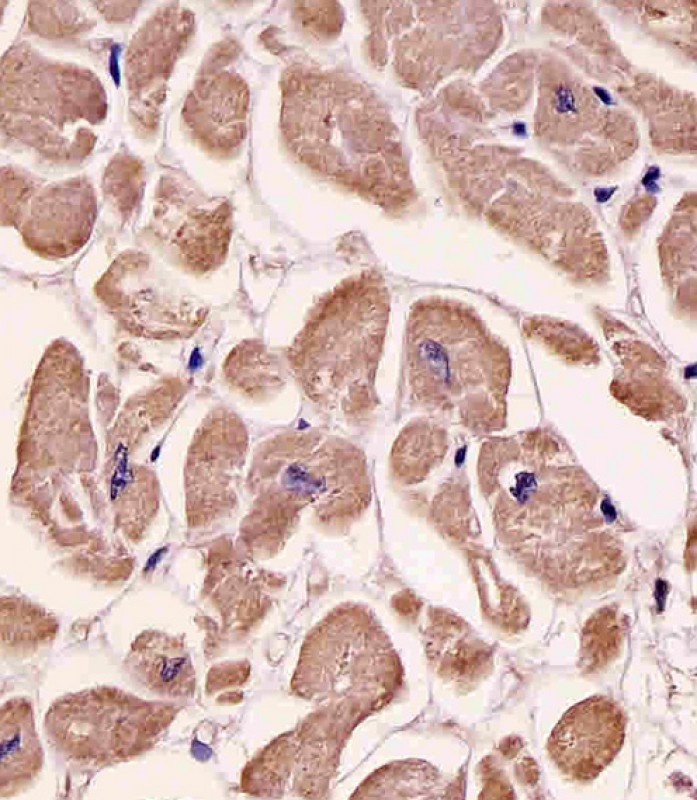
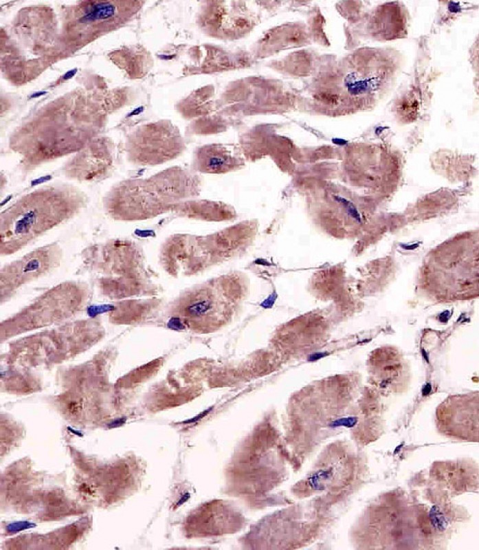
This product is a high quality SPHK1 antibody (Center).
|
||
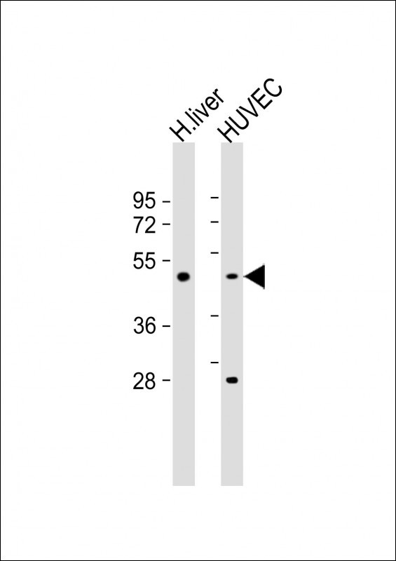
Molecular Weight |
42518 Da
|
||
Cellular Localization |
Antigen Cellular Localization:
Cytoplasm. Nucleus. Cell membrane. Note=Translocated from the cytoplasm to the plasma membrane in a CIB1-dependent manner
|
||
Host |
Rabbit
|
||
Species Reactivity |
Human, Mouse, Rat
|
||
Immunogen |
286-315 aa
|
||
Target |
This SPHK1 antibody is generated from rabbits immunized with a KLH conjugated synthetic peptide between 286-315 amino acids from the Central region of human SPHK1.
|
||
Isotype |
Rabbit Ig
|
||
Symbol |
SPHK, SPK
|
||
GeneID |
|||
UniProt ID |
|||
Function |
Catalyzes the phosphorylation of sphingosine to form sphingosine 1-phosphate (SPP), a lipid mediator with both intra- and extracellular functions. Also acts on D-erythro-sphingosine and to a lesser extent sphinganine, but not other lipids, such as D,L-threo-dihydrosphingosine, N,N-dimethylsphingosine, diacylglycerol, ceramide, or phosphatidylinositol.
|
||
Summary |
Sphingosine Kinase (SphK) catalyzes the phosphorylation of the lipid sphingosine, creating the bioactive lipid sphingosine-1-phosphate (S1P). S1P subsequently signals through cell surface G protein-coupled receptors, as well as intracellularly, to modulate cell proliferation, survival, motility and differentiation. SphK is an important signaling enzyme which is activated by diverse agents, including growth factors that signal through receptor tyrosine kinases, agents activating G protein-coupled receptors, and immunoglobulin receptors. Two SphK isotypes, SphK-1 and SphK-2, have been cloned, and both isotypes are ubiquitously expressed. SphK-1 has been shown to mediate cell growth, prevention of apoptosis, and cellular transformation, and is upregulated in a variety of human tumors. In contrast, SphK-2 increases apoptosis, and may be responsible for phosphorylating and activating the immunosuppressive drug FTY720.
|
||
Form |
Purified polyclonal antibody supplied in PBS with 0.09% (W/V) sodium azide. This antibody is purified through a protein A column, followed by peptide affinity purification. |
||
Storage & Stability |
Store at +4°C short term. For long-term storage, aliquot and store at -20°C or below. Stable for 12 months at -20°C. Avoid repeated freeze-thaw cycles.
|
||
Applications |
IHC-P, WB, E
|
||
Dilution |
IHC-P~~1:25
WB~~1:1000
|
||
Synonyms |
Sphingosine kinase 1, SK 1, SPK 1, SPHK1, SPHK, SPK
|
||
Images |
|
||
Specification |
|||
Quantity |
|
||
| Select | Brand | Catalog No. | Product Name | Pack Size | Type | Field of Research | Specification | Quantity | Price(USD) | |
| 1 | Leading Biology | APR03440G | ITGA11 Antibody (N-term) | 100 μl | Polyclonal Antibodies |
|
$495.00 | Add Ask | ||
| 2 | Leading Biology | APR04537G | CMIP Antibody (C-term) | 100 μl | Polyclonal Antibodies |
|
$495.00 | Add Ask | ||
| 3 | Leading Biology | APR12422G | Human H4 Histamine Receptor (extracellular) Antibody | 50 μl | Polyclonal Antibodies |
|
$695.00 | Add Ask | ||
| 4 | Leading Biology | APR03844G | UBE2W Antibody (C-term) | 100 μl | Polyclonal Antibodies |
|
$495.00 | Add Ask | ||
| 5 | Leading Biology | APR04349G | HECTD2 Antibody (N-term) | 100 μl | Polyclonal Antibodies |
|
$495.00 | Add Ask | ||
| 6 | Leading Biology | APR03502G | IGHG1 Antibody (Center) | 100 μl | Polyclonal Antibodies |
|
$495.00 | Add Ask |

You have 0 item in your cart
You have 0 item in your inquiry list