> Antigen, Antibodies, ELISA, Western Blot > Primary Antibody > Polyclonal Antibodies > Anoctamin-2 (extracellular) AntibodyBrand |
Leading Biology | Catalog Number |
APR11416G |
Product Type |
Polyclonal Antibodies | Field of Research |
|
Product Overview |
We constantly strive to ensure we provide our customers with the best antibodies. As a result of this work we offer this antibody in purified format.
We are in the process of updating our datasheets. If you have any questions regarding this update, please feel free to contact our technical support team.
This product is a high quality Anoctamin-2 (extracellular) antibody.
|
||
Molecular Weight |
114134 Da
|
||
Host |
Rabbit
|
||
Species Reactivity |
Mouse, Rat
|
||
Target |
Peptide (C)HSKRPEQWDLDHSLE, corresponding to amino acid residues 632-646 of mouse Anoctamin-2 (Accession Q8CFW1). 3rd extracellular loop.
|
||
GeneID |
|||
UniProt ID |
|||
Summary |
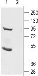
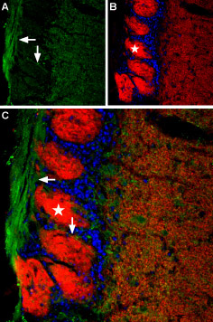
Anoctamin (ANO, or TMEM16) is a family of membrane proteins which includes 10 members. This family is named so because these channels selective to ANions and have eight (OCT) transmembrane domains. Also, these channels are subject to glycosylation in their extracellular loops and have both intracellular N- and C-termini1. Members of this family are expressed in a broad range of different organisms ranging from mammals, flies, worms, plants as well as yeast1. Alternative splicing is known to affect these channels and regarding their oligomerization state, homedimerization has been observed although when heterologously expressed, these channels may hetero oligomerize2.Ano1 (or TMEM16A, DOG1 and others) the first member to be identified was found to be a Ca2+-activated Cl- channel3-5 therefore other members are likely to also be Cl- channels. These channels are expressed in many different tissues: bronchiolar epithelial cells, pancreatic acinar cells, proximal kidney tubule epithelium, retina, dorsal root ganglia and submandibular gland1. In fact, Ano1 gained a lot of attention as its activation may serve as a therapeutic treatment for cystic fibrosis since it is also expressed in the airways6. These Ca2+-activated Cl- channels are believed to play a role in development as knock out of Ano1 in mice causes abnormal development of the trachea7. Ano2 (TMEM16B) has been shown to mediate Ca2+-activated Cl- current in olfactory epithelium and photoreceptor synapses2,8,9.Although relatively newly discovered channels, they are being discovered in many medical indications. Ano1 has become a marker in gastrointestinal tumors as its expression is significantly upregulated in such tumors10,11. Similarly, Ano1 is also highly expressed other carcinomas12,13.Abgent is pleased to offer a highly specific antibody directed against an extracellular epitope of mouse Anoctamin 2 channel. Anti-Anoctamin-2 (extracellular) antibody (#AG1307) can be used in western blot and immunohistochemical applications and has been designed to recognize Anoctamin-2 channel from mouse, rat and human samples.
|
||
Form |
Affinity purified antibody, Liquid |
||
Storage & Stability |
Store at +4°C short term. For long-term storage, aliquot and store at -20°C or below. Stable for 12 months at -20°C. Avoid repeated freeze-thaw cycles.
|
||
Applications |
WB
|
||
Dilution |
WB~~1:200-1:2000
IHC~~1:200
|
||
Synonyms |
Anoctamin-2, Ano2 {ECO:0000250|UniProtKB:Q9NQ90}
|
||
Images |
|
||
Specification |
|||
Quantity |
|
||
| Select | Brand | Catalog No. | Product Name | Pack Size | Type | Field of Research | Specification | Quantity | Price(USD) | |
| 1 | Leading Biology | APR03440G | ITGA11 Antibody (N-term) | 100 μl | Polyclonal Antibodies |
|
$495.00 | Add Ask | ||
| 2 | Leading Biology | APR04537G | CMIP Antibody (C-term) | 100 μl | Polyclonal Antibodies |
|
$495.00 | Add Ask | ||
| 3 | Leading Biology | APR12422G | Human H4 Histamine Receptor (extracellular) Antibody | 50 μl | Polyclonal Antibodies |
|
$695.00 | Add Ask | ||
| 4 | Leading Biology | APR03844G | UBE2W Antibody (C-term) | 100 μl | Polyclonal Antibodies |
|
$495.00 | Add Ask | ||
| 5 | Leading Biology | APR04349G | HECTD2 Antibody (N-term) | 100 μl | Polyclonal Antibodies |
|
$495.00 | Add Ask | ||
| 6 | Leading Biology | APR03502G | IGHG1 Antibody (Center) | 100 μl | Polyclonal Antibodies |
|
$495.00 | Add Ask |

You have 0 item in your cart
You have 0 item in your inquiry list