> Antigen, Antibodies, ELISA, Western Blot > Primary Antibody > Polyclonal Antibodies > mGluR2 (extracellular) AntibodyBrand |
Leading Biology | Catalog Number |
AMM06391G |
Product Type |
Polyclonal Antibodies | Field of Research |
|
Product Overview |
We constantly strive to ensure we provide our customers with the best antibodies. As a result of this work we offer this antibody in purified format.
We are in the process of updating our datasheets. If you have any questions regarding this update, please feel free to contact our technical support team.
This product is a high quality mGluR2 (extracellular) antibody.
|
||
Molecular Weight |
95774 Da
|
||
Host |
Rabbit
|
||
Species Reactivity |
Human, Mouse, Rat
|
||
Target |
Peptide SLSRGADGSRHIC, corresponding to amino acids 109-121 of rat mGluR2 (Accession P31421). Extracellular, N-terminus.
|
||
GeneID |
|||
UniProt ID |
|||
Summary |
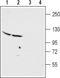
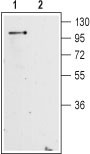
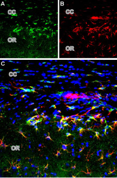
L-Glutamate, the major excitatory neurotransmitter in the central nervous system, operates through several receptors that are categorized as ionotropic (ligand-gated cation channels) or metabotropic (G-protein coupled receptors). The metabotropic glutamate receptors family includes eight members (mGluR1-8) that have been divided into three groups based on their sequence homology, pharmacology and signal transduction.Group II of the metabotropic glutamate receptors includes the mGluR2 and mGluR3 receptors. The receptors present the typical G-protein coupled receptor (GPCR) signature topology: seven transmembrane domains with a large extracellular N-terminus domain that contains the glutamate binding site, and an intracellular C-terminus one.1,2 mGluR2 and mGluR3 are coupled to Gi/Go and hence inhibit cAMP formation following receptor activation.1, 2 mGluR2 is widely distributed throughout the brain with high expression in several limbic areas including the cortex, hippocampus and amygdala. mGluR2 is localized primarily presinaptically, although postsynaptical localization has also been described.In line with its presynaptical localization, mGluR2 is thought to function as an autoreceptor in a negative feedback mechanism that suppress further release of glutamate from the cell on which it is expressed. The involvement of mGluR2 in neuronal excitability and synaptic transmission suggests that modulation of this receptor is a promising strategy for the treatment of neurological and neuropsychiatric disorders such as anxiety, schizophrenia, and pain.3,4 Abgent is pleased to offer a highly specific antibody directed against an epitope located at the extracellular N-terminus domain of the rat mGluR2 receptor. The epitope is specific for mGluR2 and will not cross-react with the closely related mGluR3 channel. Anti- mGluR2 (extracellular) antibody (#AG1258) can be used in Western blot analysis and immunohistochemical applications, and recognizes mGluR2 from rat and mouse samples.
|
||
Form |
Affinity purified antibody, Liquid |
||
Storage & Stability |
Store at +4°C short term. For long-term storage, aliquot and store at -20°C or below. Stable for 12 months at -20°C. Avoid repeated freeze-thaw cycles.
|
||
Applications |
WB, IHC
|
||
Dilution |
WB~~1:200-1:2000
|
||
Synonyms |
Metabotropic glutamate receptor 2, mGluR2, Grm2, Gprc1b, Mglur2
|
||
Images |
|
||
Specification |
|||
Quantity |
|
||
| Select | Brand | Catalog No. | Product Name | Pack Size | Type | Field of Research | Specification | Quantity | Price(USD) | |
| 1 | Leading Biology | APR03440G | ITGA11 Antibody (N-term) | 100 μl | Polyclonal Antibodies |
|
$495.00 | Add Ask | ||
| 2 | Leading Biology | APR04537G | CMIP Antibody (C-term) | 100 μl | Polyclonal Antibodies |
|
$495.00 | Add Ask | ||
| 3 | Leading Biology | APR12422G | Human H4 Histamine Receptor (extracellular) Antibody | 50 μl | Polyclonal Antibodies |
|
$695.00 | Add Ask | ||
| 4 | Leading Biology | APR03844G | UBE2W Antibody (C-term) | 100 μl | Polyclonal Antibodies |
|
$495.00 | Add Ask | ||
| 5 | Leading Biology | APR04349G | HECTD2 Antibody (N-term) | 100 μl | Polyclonal Antibodies |
|
$495.00 | Add Ask | ||
| 6 | Leading Biology | APR03502G | IGHG1 Antibody (Center) | 100 μl | Polyclonal Antibodies |
|
$495.00 | Add Ask |

You have 0 item in your cart
You have 0 item in your inquiry list