> Antigen, Antibodies, ELISA, Western Blot > Primary Antibody > Polyclonal Antibodies > STIM1 (extracellular) AntibodyBrand |
Leading Biology | Catalog Number |
APR10299G |
Product Type |
Polyclonal Antibodies | Field of Research |
|
Product Overview |
We constantly strive to ensure we provide our customers with the best antibodies. As a result of this work we offer this antibody in purified format.
We are in the process of updating our datasheets. If you have any questions regarding this update, please feel free to contact our technical support team.
This product is a high quality STIM1 (extracellular) antibody.
|
||
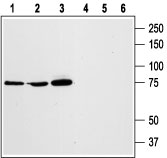
Molecular Weight |
77423 Da
|
||
Host |
Rabbit
|
||
Species Reactivity |
Human, Mouse, Rat
|
||
Target |
Peptide CHSEDEKLSFEAVR, corresponding to amino acid residues 56-69 of human STIM1 (Accession Q13586).???? STIM1 expressed on the plasma membrane: Extracellular, N-terminus. STIM1 expressed on the ER: Luminal, N-terminus.
|
||
GeneID |
|||
UniProt ID |
|||
Summary |
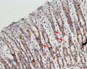
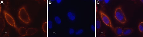
Cytosolic Ca2+ has long been known to act as a key second messenger in many intracellular pathways including synaptic transmission, muscle contraction, hormonal secretion, and cell growth and proliferation.1,2 The mechanism controlling the influx of intracellular Ca2+ either by calcium-release-activated Ca2+ channels (CRAC) or from intracellular stores has lately become of great interest.Recently, several key players of the store-operated complex have been identified.3 The Orai family consists of three members, Orai1-3, and the STIM family, which consists of two members, STIM1 and STIM2. Orai1 (also known as CRACM1) acts as the store-operated calcium channel (SOC) and STIM1 as the endoplasmic reticulum (ER) Ca2+ sensor.3,4 The majority of STIM1 appears to be localized intracellularly at the ER membrane while low expression of STIM1 has been detected on the cell surface of several cell types.5 STIM1 has an amino-terminal EF hand Ca2+ -binding domain facing the lumen of the ER.6 Upon Ca2+ store depletion, STIM1 molecules are redistributed in punctae underneath the plasma membrane and activate SOCs.Several possible interactions between STIM1 and Orai1 have been suggested. The most simple and cited is a dynamic interaction between the cytosolic C-terminus of STIM1 and the cytoplasmic domain of the Orai1 channel.7-9 STIM1 is assumed to regulate the activity of all known SOCs, including native SOCs.5 Consistent with their important role as calcium sensors within the ER, STIM1 proteins are ubiquitously expressed.Abgent is pleased to offer a highly specific antibody directed against an extracellular epitope of human STIM1. Anti-STIM1 (extracellular) antibody (#AG1335) can be used in western blot, immunocytochemical, immunohistochemical, and indirect flow cytometry applications and has been designed torecognize STIM1 from human, rat, mouse, bovine, and canis samples.
|
||
Form |
Affinity purified antibody, Liquid |
||
Storage & Stability |
Store at +4°C short term. For long-term storage, aliquot and store at -20°C or below. Stable for 12 months at -20°C. Avoid repeated freeze-thaw cycles.
|
||
Applications |
WB, LCI
|
||
Dilution |
WB~~1:200-1:2000
IHC~~1:100
ICC~~1:50
|
||
Synonyms |
Stromal interaction molecule 1, STIM1, GOK
|
||
Images |
|
||
Specification |
|||
Quantity |
|
||
| Select | Brand | Catalog No. | Product Name | Pack Size | Type | Field of Research | Specification | Quantity | Price(USD) | |
| 1 | Leading Biology | APR03440G | ITGA11 Antibody (N-term) | 100 μl | Polyclonal Antibodies |
|
$495.00 | Add Ask | ||
| 2 | Leading Biology | APR04537G | CMIP Antibody (C-term) | 100 μl | Polyclonal Antibodies |
|
$495.00 | Add Ask | ||
| 3 | Leading Biology | APR12422G | Human H4 Histamine Receptor (extracellular) Antibody | 50 μl | Polyclonal Antibodies |
|
$695.00 | Add Ask | ||
| 4 | Leading Biology | APR03844G | UBE2W Antibody (C-term) | 100 μl | Polyclonal Antibodies |
|
$495.00 | Add Ask | ||
| 5 | Leading Biology | APR04349G | HECTD2 Antibody (N-term) | 100 μl | Polyclonal Antibodies |
|
$495.00 | Add Ask | ||
| 6 | Leading Biology | APR03502G | IGHG1 Antibody (Center) | 100 μl | Polyclonal Antibodies |
|
$495.00 | Add Ask |

You have 0 item in your cart
You have 0 item in your inquiry list