> Antigen, Antibodies, ELISA, Western Blot > Primary Antibody > Polyclonal Antibodies > Cleaved LC3A AntibodyBrand |
Leading Biology | Catalog Number |
APR26972G |
Product Type |
Polyclonal Antibodies | Field of Research |
|
Product Overview |
We constantly strive to ensure we provide our customers with the best antibodies. As a result of this work we offer this antibody in purified format.
We are in the process of updating our datasheets. If you have any questions regarding this update, please feel free to contact our technical support team.
This product is a high quality Cleaved LC3A antibody.
|
||
Molecular Weight |
14272 Da
|
||
Cellular Localization |
Antigen Cellular Localization:
Cytoplasm, cytoskeleton. Endomembrane system; Lipid-anchor. Cytoplasmic vesicle, autophagosome membrane; Lipid-anchor. Note=LC3-II binds to the autophagic membranes
|
||
Host |
Rabbit
|
||
Species Reactivity |
Human, Mouse
|
||
Immunogen |
89-120 aa
|
||
Target |
This Cleaved LC3A antibody is generated from rabbits immunized with a KLH conjugated synthetic peptide between 89-120 amino acids from human Cleaved LC3A.
|
||
Isotype |
Rabbit Ig
|
||
GeneID |
|||
UniProt ID |
|||
Function |
Ubiquitin-like modifier involved in formation of autophagosomal vacuoles (autophagosomes). Whereas LC3s are involved in elongation of the phagophore membrane, the GABARAP/GATE-16 subfamily is essential for a later stage in autophagosome maturation.
|
||
Summary |
Macroautophagy is the major inducible pathway for the general turnover of cytoplasmic constituents in eukaryotic cells, it is also responsible for the degradation of active cytoplasmic enzymes and organelles during nutrient starvation. Macroautophagy involves the formation of double-membrane bound autophagosomes which enclose the cytoplasmic constituent targeted for degradation in a membrane bound structure, which then fuse with the lysosome (or vacuole) releasing a single-membrane bound autophagic bodies which are then degraded within the lysosome (or vacuole). MAP1A and MAP1B are microtubule-associated proteins which mediate the physical interactions between microtubules and components of the cytoskeleton. These proteins are involved in formation of autophagosomal vacuoles (autophagosomes). MAP1A and MAP1B each consist of a heavy chain subunit and multiple light chain subunits. MAP1LC3a is one of the light chain subunits and can associate with either MAP1A or MAP1B. The precursor molecule is cleaved by APG4B/ATG4B to form the cytosolic form, LC3-I. This is activated by APG7L/ATG7, transferred to ATG3 and conjugated to phospholipid to form the membrane-bound form, LC3-II.
|
||
Form |
Purified polyclonal antibody supplied in PBS with 0.09% (W/V) sodium azide. This antibody is purified through a protein A column, followed by peptide affinity purification. |
||
Storage & Stability |
Store at +4°C short term. For long-term storage, aliquot and store at -20°C or below. Stable for 12 months at -20°C. Avoid repeated freeze-thaw cycles.
|
||
Applications |
WB, IHC-P, IF, ICC, E
|
||
Dilution |
IF~~1:25
IHC-P~~1:10~50
WB~~1:1000
ICC~~1:10~50
|
||
Synonyms |
Microtubule-associated proteins 1A/1B light chain 3A, Autophagy-related protein LC3 A, Autophagy-related ubiquitin-like modifier LC3 A, MAP1 light chain 3-like protein 1, MAP1A/MAP1B light chain 3 A, MAP1A/MAP1B LC3 A, Microtubule-associated protein 1 light chain 3 alpha, MAP1LC3A
|
||
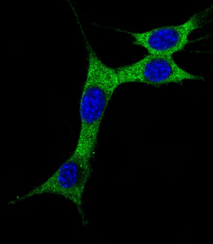
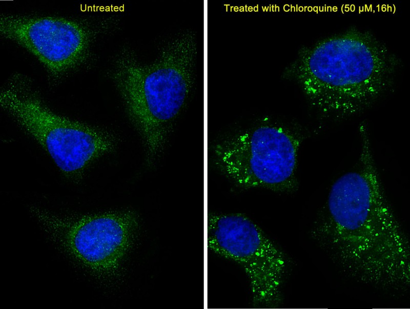
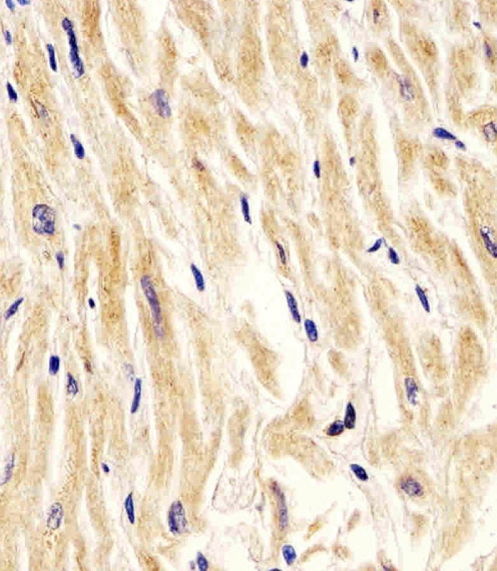
Images |
|
||
Specification |
|||
Quantity |
|
||
| Select | Brand | Catalog No. | Product Name | Pack Size | Type | Field of Research | Specification | Quantity | Price(USD) | |
| 1 | Leading Biology | APR03440G | ITGA11 Antibody (N-term) | 100 μl | Polyclonal Antibodies |
|
$495.00 | Add Ask | ||
| 2 | Leading Biology | APR04537G | CMIP Antibody (C-term) | 100 μl | Polyclonal Antibodies |
|
$495.00 | Add Ask | ||
| 3 | Leading Biology | APR12422G | Human H4 Histamine Receptor (extracellular) Antibody | 50 μl | Polyclonal Antibodies |
|
$695.00 | Add Ask | ||
| 4 | Leading Biology | APR03844G | UBE2W Antibody (C-term) | 100 μl | Polyclonal Antibodies |
|
$495.00 | Add Ask | ||
| 5 | Leading Biology | APR04349G | HECTD2 Antibody (N-term) | 100 μl | Polyclonal Antibodies |
|
$495.00 | Add Ask | ||
| 6 | Leading Biology | APR03502G | IGHG1 Antibody (Center) | 100 μl | Polyclonal Antibodies |
|
$495.00 | Add Ask |

You have 0 item in your cart
You have 0 item in your inquiry list