> Antigen, Antibodies, ELISA, Western Blot > Primary Antibody > Polyclonal Antibodies > DVL1 Antibody (Center)Brand |
Leading Biology | Catalog Number |
AMM06980G |
Product Type |
Polyclonal Antibodies | Field of Research |
|
Product Overview |
We constantly strive to ensure we provide our customers with the best antibodies. As a result of this work we offer this antibody in purified format.
We are in the process of updating our datasheets. If you have any questions regarding this update, please feel free to contact our technical support team.
This product is a high quality DVL1 antibody (Center).
|
||
Molecular Weight |
75187 Da
|
||
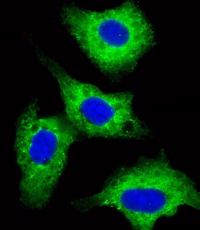
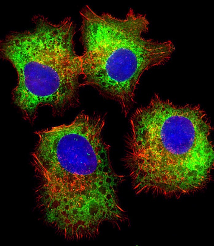
Cellular Localization |
Antigen Cellular Localization:
Cell membrane; Peripheral membrane protein; Cytoplasmic side Cytoplasm, cytosol. Cytoplasmic vesicle. Note=Localizes at the cell membrane upon interaction with frizzled family members.
|
||
Host |
Rabbit
|
||
Species Reactivity |
Human, Mouse
|
||
Immunogen |
442-470 aa
|
||
Target |
This DVL1 antibody is generated from rabbits immunized with a KLH conjugated synthetic peptide between 442-470 amino acids from the Central region of human DVL1.
|
||
Isotype |
Rabbit Ig
|
||
GeneID |
|||
UniProt ID |
|||
Function |
Participates in Wnt signaling by binding to the cytoplasmic C-terminus of frizzled family members and transducing the Wnt signal to down-stream effectors. Plays a role both in canonical and non-canonical Wnt signaling. Plays a role in the signal transduction pathways mediated by multiple Wnt genes. Required for LEF1 activation upon WNT1 and WNT3A signaling. DVL1 and PAK1 form a ternary complex with MUSK which is important for MUSK-dependent regulation of AChR clustering during the formation of the neuromuscular junction (NMJ).
|
||
Summary |
DVL1, the human homolog of the Drosophila dishevelled gene(dsh) encodes a cytoplasmic phosphoprotein that regulates cellproliferation, acting as a transducer molecule for developmentalprocesses, including segmentation and neuroblast specification.DVL1 is a candidate gene for neuroblastomatous transformation. TheSchwartz-Jampel syndrome and Charcot-Marie-Tooth disease type 2Ahave been mapped to the same region as DVL1. The phenotypes ofthese diseases may be consistent with defects which might beexpected from aberrant expression of a DVL gene during development.
|
||
Form |
Purified polyclonal antibody supplied in PBS with 0.09% (W/V) sodium azide. This antibody is purified through a protein A column, followed by peptide affinity purification. |
||
Storage & Stability |
Store at +4°C short term. For long-term storage, aliquot and store at -20°C or below. Stable for 12 months at -20°C. Avoid repeated freeze-thaw cycles.
|
||
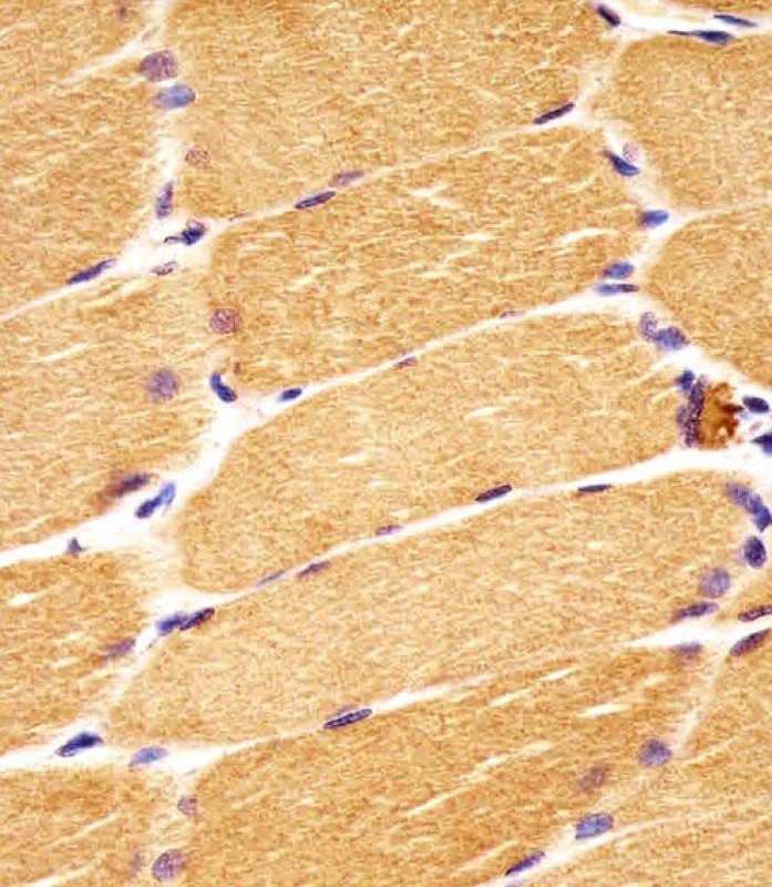
Applications |
IF, IHC-P, WB, E
|
||
Dilution |
IF~~1:10~50
IHC-P~~1:10~50
WB~~1:1000
|
||
Synonyms |
Segment polarity protein dishevelled homolog DVL-1, Dishevelled-1, DSH homolog 1, DVL1
|
||
Images |
|
||
Specification |
|||
Quantity |
|
||
| Select | Brand | Catalog No. | Product Name | Pack Size | Type | Field of Research | Specification | Quantity | Price(USD) | |
| 1 | Leading Biology | APR03440G | ITGA11 Antibody (N-term) | 100 μl | Polyclonal Antibodies |
|
$495.00 | Add Ask | ||
| 2 | Leading Biology | APR04537G | CMIP Antibody (C-term) | 100 μl | Polyclonal Antibodies |
|
$495.00 | Add Ask | ||
| 3 | Leading Biology | APR12422G | Human H4 Histamine Receptor (extracellular) Antibody | 50 μl | Polyclonal Antibodies |
|
$695.00 | Add Ask | ||
| 4 | Leading Biology | APR03844G | UBE2W Antibody (C-term) | 100 μl | Polyclonal Antibodies |
|
$495.00 | Add Ask | ||
| 5 | Leading Biology | APR04349G | HECTD2 Antibody (N-term) | 100 μl | Polyclonal Antibodies |
|
$495.00 | Add Ask | ||
| 6 | Leading Biology | APR03502G | IGHG1 Antibody (Center) | 100 μl | Polyclonal Antibodies |
|
$495.00 | Add Ask |

You have 0 item in your cart
You have 0 item in your inquiry list