> Antigen, Antibodies, ELISA, Western Blot > Primary Antibody > Polyclonal Antibodies > CKIP-1 Antibody (N-term)Brand |
Leading Biology | Catalog Number |
APR15459G |
Product Type |
Polyclonal Antibodies | Field of Research |
|
Product Overview |
We constantly strive to ensure we provide our customers with the best antibodies. As a result of this work we offer this antibody in purified format.
We are in the process of updating our datasheets. If you have any questions regarding this update, please feel free to contact our technical support team.
This product is a high quality CKIP-1 antibody (N-term).
|
||
Molecular Weight |
46237 Da
|
||
Cellular Localization |
Antigen Cellular Localization:
Cell membrane. Nucleus. Cytoplasm. Note=Predominantly localized to the plasma membrane. In C2C12 cells, with the absence of growth factor, it is found in the nucleus. It rapidly translocates to the plasma membrane after insulin stimulation. In response to TNF, it translocates from the plasma membrane to the cytoplasm and then to the nucleus accompanied by cleavage by caspase-3. However, the subcellular location is highly dependent of the cell type, and this explains why it is found exclusively at the plasma membrane, in some type of cells
|
||
Host |
Rabbit
|
||
Species Reactivity |
Human
|
||
Immunogen |
49-77 aa
|
||
Target |
This CKIP-1 antibody is generated from rabbits immunized with a KLH conjugated synthetic peptide between 49-77 amino acids from the N-terminal region of human CKIP-1.
|
||
Isotype |
Rabbit Ig
|
||
Symbol |
CKIP1, OC120
|
||
GeneID |
|||
UniProt ID |
|||
Function |
Plays a role in the regulation of the actin cytoskeleton through its interactions with actin capping protein (CP). May function to target CK2 to the plasma membrane thereby serving as an adapter to facilitate the phosphorylation of CP by protein kinase 2 (CK2). Appears to target ATM to the plasma membrane. Appears to also inhibit tumor cell growth by inhibiting AKT- mediated cell-survival. Also implicated in PI3K-regulated muscle differentiation, the regulation of AP-1 activity (plasma membrane bound AP-1 regulator that translocates to the nucleus) and the promotion of apoptosis induced by tumor necrosis factor TNF. When bound to PKB, it inhibits it probably by decreasing PKB level of phosphorylation.
|
||
Summary |
CKIP-1 plays a role in the regulation of the actin cytoskeleton through its interactions with actin capping protein (CP). This protein may function to target CK2 to the plasma membrane thereby serving as an adapter to facilitate the phosphorylation of CP by protein kinase 2 (CK2). It appears to target ATM to the plasma membrane and appears to also inhibit tumor cell growth by inhibiting AKT-mediated cell-survival. Also implicated in PI3K-regulated muscle differentiation, the regulation of AP-1 activity (plasma membrane bound AP-1 regulator that translocates to the nucleus) and the promotion of apoptosis induced by tumor necrosis factor TNF. When bound to PKB, it inhibits it probably by decreasing PKB level of phosphorylation.
|
||
Form |
Purified polyclonal antibody supplied in PBS with 0.09% (W/V) sodium azide. This antibody is prepared by Saturated Ammonium Sulfate (SAS) precipitation followed by dialysis against PBS. |
||
Storage & Stability |
Store at +4°C short term. For long-term storage, aliquot and store at -20°C or below. Stable for 12 months at -20°C. Avoid repeated freeze-thaw cycles.
|
||
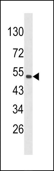
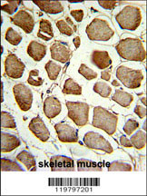
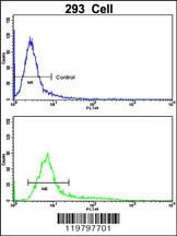
Applications |
WB, IHC-P, FC, E
|
||
Dilution |
WB~~1:1000
IHC-P~~1:50~100
FC~~1:10~50
|
||
Synonyms |
Pleckstrin homology domain-containing family O member 1, PH domain-containing family O member 1, C-Jun-binding protein, JBP, Casein kinase 2-interacting protein 1, CK2-interacting protein 1, CKIP-1, Osteoclast maturation-associated gene 120 protein, PLEKHO1, CKIP1, OC120
|
||
Images |
|
||
Specification |
|||
Quantity |
|
||
| Select | Brand | Catalog No. | Product Name | Pack Size | Type | Field of Research | Specification | Quantity | Price(USD) | |
| 1 | Leading Biology | APR03440G | ITGA11 Antibody (N-term) | 100 μl | Polyclonal Antibodies |
|
$495.00 | Add Ask | ||
| 2 | Leading Biology | APR04537G | CMIP Antibody (C-term) | 100 μl | Polyclonal Antibodies |
|
$495.00 | Add Ask | ||
| 3 | Leading Biology | APR12422G | Human H4 Histamine Receptor (extracellular) Antibody | 50 μl | Polyclonal Antibodies |
|
$695.00 | Add Ask | ||
| 4 | Leading Biology | APR03844G | UBE2W Antibody (C-term) | 100 μl | Polyclonal Antibodies |
|
$495.00 | Add Ask | ||
| 5 | Leading Biology | APR04349G | HECTD2 Antibody (N-term) | 100 μl | Polyclonal Antibodies |
|
$495.00 | Add Ask | ||
| 6 | Leading Biology | APR03502G | IGHG1 Antibody (Center) | 100 μl | Polyclonal Antibodies |
|
$495.00 | Add Ask |

You have 0 item in your cart
You have 0 item in your inquiry list