> Antigen, Antibodies, ELISA, Western Blot > Primary Antibody > Polyclonal Antibodies > FLT3 (CD135) Antibody (C-term)Brand |
Leading Biology | Catalog Number |
APR16007G |
Product Type |
Polyclonal Antibodies | Field of Research |
|
Product Overview |
We constantly strive to ensure we provide our customers with the best antibodies. As a result of this work we offer this antibody in purified format.
We are in the process of updating our datasheets. If you have any questions regarding this update, please feel free to contact our technical support team.
This product is a high quality FLT3 (CD135) antibody (C-term).
|
||
Molecular Weight |
112903 Da
|
||
Cellular Localization |
Antigen Cellular Localization:
Membrane; Single-pass type I membrane protein. Endoplasmic reticulum lumen. Note=Constitutively activated mutant forms with internal tandem duplications are less efficiently transported to the cell surface and a significant proportion is retained in an immature form in the endoplasmic reticulum lumen. The activated kinase is rapidly targeted for degradation
|
||
Host |
Rabbit
|
||
Species Reactivity |
Human
|
||
Immunogen |
951-982 aa
|
||
Target |
This FLT3 (CD135) antibody is generated from rabbits immunized with a KLH conjugated synthetic peptide between 951-982 amino acids from the C-terminal region of human FLT3 (CD135).
|
||
Isotype |
Rabbit Ig
|
||
Symbol |
CD135, FLK2, STK1
|
||
GeneID |
|||
UniProt ID |
|||
Function |
Tyrosine-protein kinase that acts as cell-surface receptor for the cytokine FLT3LG and regulates differentiation, proliferation and survival of hematopoietic progenitor cells and of dendritic cells. Promotes phosphorylation of SHC1 and AKT1, and activation of the downstream effector MTOR. Promotes activation of RAS signaling and phosphorylation of downstream kinases, including MAPK1/ERK2 and/or MAPK3/ERK1. Promotes phosphorylation of FES, FER, PTPN6/SHP, PTPN11/SHP-2, PLCG1, and STAT5A and/or STAT5B. Activation of wild-type FLT3 causes only marginal activation of STAT5A or STAT5B. Mutations that cause constitutive kinase activity promote cell proliferation and resistance to apoptosis via the activation of multiple signaling pathways.
|
||
Summary |
Protein kinases are enzymes that transfer a phosphate group from a phosphate donor, generally the g phosphate of ATP, onto an acceptor amino acid in a substrate protein. By this basic mechanism, protein kinases mediate most of the signal transduction in eukaryotic cells, regulating cellular metabolism, transcription, cell cycle progression, cytoskeletal rearrangement and cell movement, apoptosis, and differentiation. With more than 500 gene products, the protein kinase family is one of the largest families of proteins in eukaryotes. The family has been classified in 8 major groups based on sequence comparison of their tyrosine (PTK) or serine/threonine (STK) kinase catalytic domains. The tyrosine kinase (TK) group is mainly involved in the regulation of cell-cell interactions such as differentiation, adhesion, motility and death. There are currently about 90 TK genes sequenced, 58 are of receptor protein TK (e.g. EGFR, EPH, FGFR, PDGFR, TRK, and VEGFR families), and 32 of cytosolic TK (e.g. ABL, FAK, JAK, and SRC families).
|
||
Form |
Purified polyclonal antibody supplied in PBS with 0.09% (W/V) sodium azide. This antibody is prepared by Saturated Ammonium Sulfate (SAS) precipitation followed by dialysis against PBS. |
||
Storage & Stability |
Store at +4°C short term. For long-term storage, aliquot and store at -20°C or below. Stable for 12 months at -20°C. Avoid repeated freeze-thaw cycles.
|
||
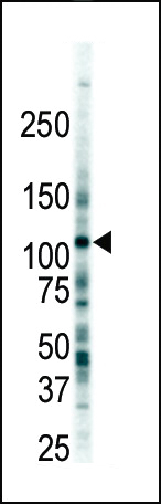
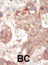
Applications |
WB, IHC-P, E
|
||
Dilution |
WB~~1:1000
IHC-P~~1:50~100
|
||
Synonyms |
Receptor-type tyrosine-protein kinase FLT3, FL cytokine receptor, Fetal liver kinase-2, FLK-2, Fms-like tyrosine kinase 3, FLT-3, Stem cell tyrosine kinase 1, STK-1, CD135, FLT3, CD135, FLK2, STK1
|
||
Images |
|
||
Specification |
|||
Quantity |
|
||
| Select | Brand | Catalog No. | Product Name | Pack Size | Type | Field of Research | Specification | Quantity | Price(USD) | |
| 1 | Leading Biology | APR03440G | ITGA11 Antibody (N-term) | 100 μl | Polyclonal Antibodies |
|
$495.00 | Add Ask | ||
| 2 | Leading Biology | APR04537G | CMIP Antibody (C-term) | 100 μl | Polyclonal Antibodies |
|
$495.00 | Add Ask | ||
| 3 | Leading Biology | APR12422G | Human H4 Histamine Receptor (extracellular) Antibody | 50 μl | Polyclonal Antibodies |
|
$695.00 | Add Ask | ||
| 4 | Leading Biology | APR03844G | UBE2W Antibody (C-term) | 100 μl | Polyclonal Antibodies |
|
$495.00 | Add Ask | ||
| 5 | Leading Biology | APR04349G | HECTD2 Antibody (N-term) | 100 μl | Polyclonal Antibodies |
|
$495.00 | Add Ask | ||
| 6 | Leading Biology | APR03502G | IGHG1 Antibody (Center) | 100 μl | Polyclonal Antibodies |
|
$495.00 | Add Ask |

You have 0 item in your cart
You have 0 item in your inquiry list