> Antigen, Antibodies, ELISA, Western Blot > Primary Antibody > Polyclonal Antibodies > PKC nu AntibodyBrand |
Leading Biology | Catalog Number |
APR14376G |
Product Type |
Polyclonal Antibodies | Field of Research |
|
Product Overview |
We constantly strive to ensure we provide our customers with the best antibodies. As a result of this work we offer this antibody in purified format.
We are in the process of updating our datasheets. If you have any questions regarding this update, please feel free to contact our technical support team.
This product is a high quality PKC nu antibody.
|
||
Molecular Weight |
100471 Da
|
||
Cellular Localization |
Antigen Cellular Localization:
Cytoplasm. Membrane. Note=Translocation to the cell membrane is required for kinase activation
|
||
Host |
Rabbit
|
||
Species Reactivity |
Human
|
||
Immunogen |
352-384 aa
|
||
Target |
This PKC-nu antibody is generated from rabbits immunized with a KLH conjugated synthetic peptide between 352-384 amino acids from human PKC-nu.
|
||
Isotype |
Rabbit Ig
|
||
Symbol |
EPK2, PRKCN
|
||
GeneID |
|||
UniProt ID |
|||
Function |
Converts transient diacylglycerol (DAG) signals into prolonged physiological effects, downstream of PKC. Involved in resistance to oxidative stress (By similarity).
|
||
Summary |
Protein kinase C (PKC) is a family of serine- and threonine-specific protein kinases that can be activated by calcium and second messenger diacylglycerol. PKC family members phosphorylate a wide variety of protein targets and are known to be involved in diverse cellular signaling pathways. PKC also serve as major receptors for phorbol esters, a class of tumor promoters. Each member of the PKC family has a specific expression profile and is believed to play distinct roles in cells. PKC nu is one of the PKC family members. This kinase can be activated rapidly by the agonists of G protein-coupled receptors. It resides in both cytoplasm and nucleus, and its nuclear accumulation is found to be dramatically enhanced in response to its activation. This kinase can also be activated after B-cell antigen receptor (BCR) engagement, which requires intact phopholipase C gamma and the involvement of other PKC family members.
|
||
Form |
Purified polyclonal antibody supplied in PBS with 0.09% (W/V) sodium azide. This antibody is prepared by Saturated Ammonium Sulfate (SAS) precipitation followed by dialysis against PBS. |
||
Storage & Stability |
Store at +4°C short term. For long-term storage, aliquot and store at -20°C or below. Stable for 12 months at -20°C. Avoid repeated freeze-thaw cycles.
|
||
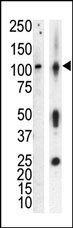
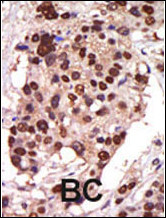
Applications |
WB, IHC-P, E
|
||
Dilution |
WB~~1:1000
IHC-P~~1:50~100
|
||
Synonyms |
Serine/threonine-protein kinase D3, Protein kinase C nu type, Protein kinase EPK2, nPKC-nu, PRKD3, EPK2, PRKCN
|
||
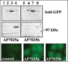
Images |
|
||
Specification |
|||
Quantity |
|
||
| Select | Brand | Catalog No. | Product Name | Pack Size | Type | Field of Research | Specification | Quantity | Price(USD) | |
| 1 | Leading Biology | APR03440G | ITGA11 Antibody (N-term) | 100 μl | Polyclonal Antibodies |
|
$495.00 | Add Ask | ||
| 2 | Leading Biology | APR04537G | CMIP Antibody (C-term) | 100 μl | Polyclonal Antibodies |
|
$495.00 | Add Ask | ||
| 3 | Leading Biology | APR12422G | Human H4 Histamine Receptor (extracellular) Antibody | 50 μl | Polyclonal Antibodies |
|
$695.00 | Add Ask | ||
| 4 | Leading Biology | APR03844G | UBE2W Antibody (C-term) | 100 μl | Polyclonal Antibodies |
|
$495.00 | Add Ask | ||
| 5 | Leading Biology | APR04349G | HECTD2 Antibody (N-term) | 100 μl | Polyclonal Antibodies |
|
$495.00 | Add Ask | ||
| 6 | Leading Biology | APR03502G | IGHG1 Antibody (Center) | 100 μl | Polyclonal Antibodies |
|
$495.00 | Add Ask |

You have 0 item in your cart
You have 0 item in your inquiry list