> Antigen, Antibodies, ELISA, Western Blot > Primary Antibody > Polyclonal Antibodies > SYVN1 (HRD1) Antibody (C-term)Brand |
Leading Biology | Catalog Number |
APR14266G |
Product Type |
Polyclonal Antibodies | Field of Research |
|
Product Overview |
We constantly strive to ensure we provide our customers with the best antibodies. As a result of this work we offer this antibody in purified format.
We are in the process of updating our datasheets. If you have any questions regarding this update, please feel free to contact our technical support team.
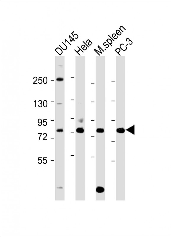
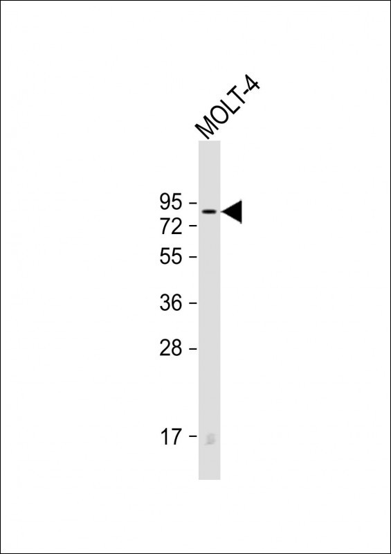
This product is a high quality SYVN1 (HRD1) antibody (C-term).
|
||
Molecular Weight |
67685 Da
|
||
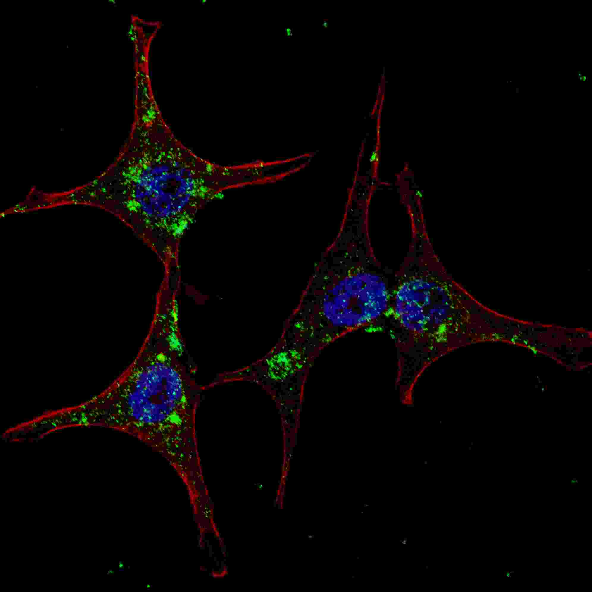
Cellular Localization |
Antigen Cellular Localization:
Endoplasmic reticulum membrane; Multi- pass membrane protein
|
||
Host |
Rabbit
|
||
Species Reactivity |
Human, Mouse
|
||
Immunogen |
586-617 aa
|
||
Target |
This SYVN1 (HRD1) antibody is generated from rabbits immunized with a KLH conjugated synthetic peptide between 586-617 amino acids from the C-terminal region of human SYVN1 (HRD1).
|
||
Isotype |
Rabbit Ig
|
||
Symbol |
HRD1, KIAA1810
|
||
GeneID |
|||
UniProt ID |
|||
Function |
Acts as an E3 ubiquitin-protein ligase which accepts ubiquitin specifically from endoplasmic reticulum-associated UBC7 E2 ligase and transfers it to substrates, promoting their degradation. Component of the endoplasmic reticulum quality control (ERQC) system also called ER-associated degradation (ERAD) involved in ubiquitin-dependent degradation of misfolded endoplasmic reticulum proteins. Also promotes the degradation of normal but naturally short-lived proteins such as SGK. Protects cells from ER stress-induced apoptosis. Protects neurons from apoptosis induced by polyglutamine-expanded huntingtin (HTT) or unfolded GPR37 by promoting their degradation. Sequesters p53/TP53 in the cytoplasm and promotes its degradation, thereby negatively regulating its biological function in transcription, cell cycle regulation and apoptosis.
|
||
Summary |
HRD1 is a ubiquitin ligase whose expression is induced by the unfolded protein response (UPR) following endoplasmic reticulum stress. Expression of HRD1 protects cells from apoptosis by inducing degradation of abnormally processed proteins that accumulate in the endoplasmic reticulum. HRD1 is expressed in many tissues, strongly expressed in brain, pancreas, liver, kidney and skeletal muscle. Amano T, et al. reported that Synoviolin/Hrd1 (expressed in rheumatoid synovium) is a novel causative factor for arthropathy by triggering synovial cell outgrowth through its antiapoptotic effects. HRD1 contains one ring-type zinc finger.
|
||
Form |
Purified polyclonal antibody supplied in PBS with 0.09% (W/V) sodium azide. This antibody is purified through a protein A column, followed by peptide affinity purification. |
||
Storage & Stability |
Store at +4°C short term. For long-term storage, aliquot and store at -20°C or below. Stable for 12 months at -20°C. Avoid repeated freeze-thaw cycles.
|
||
Applications |
WB, IHC-P, IF, E
|
||
Dilution |
WB~~1:1000
IF~~1:200
IHC-P~~1:50~100
|
||
Synonyms |
E3 ubiquitin-protein ligase synoviolin, 632-, Synovial apoptosis inhibitor 1, SYVN1, HRD1, KIAA1810
|
||
Images |
|
||
Specification |
|||
Quantity |
|
||
| Select | Brand | Catalog No. | Product Name | Pack Size | Type | Field of Research | Specification | Quantity | Price(USD) | |
| 1 | Leading Biology | APR03440G | ITGA11 Antibody (N-term) | 100 μl | Polyclonal Antibodies |
|
$495.00 | Add Ask | ||
| 2 | Leading Biology | APR04537G | CMIP Antibody (C-term) | 100 μl | Polyclonal Antibodies |
|
$495.00 | Add Ask | ||
| 3 | Leading Biology | APR12422G | Human H4 Histamine Receptor (extracellular) Antibody | 50 μl | Polyclonal Antibodies |
|
$695.00 | Add Ask | ||
| 4 | Leading Biology | APR03844G | UBE2W Antibody (C-term) | 100 μl | Polyclonal Antibodies |
|
$495.00 | Add Ask | ||
| 5 | Leading Biology | APR04349G | HECTD2 Antibody (N-term) | 100 μl | Polyclonal Antibodies |
|
$495.00 | Add Ask | ||
| 6 | Leading Biology | APR03502G | IGHG1 Antibody (Center) | 100 μl | Polyclonal Antibodies |
|
$495.00 | Add Ask |

You have 0 item in your cart
You have 0 item in your inquiry list