> Antigen, Antibodies, ELISA, Western Blot > Primary Antibody > Polyclonal Antibodies > BNIP3 Antibody (BH3 Domain Specific)Brand |
Leading Biology | Catalog Number |
APR10804G |
Product Type |
Polyclonal Antibodies | Field of Research |
|
Product Overview |
We constantly strive to ensure we provide our customers with the best antibodies. As a result of this work we offer this antibody in purified format.
We are in the process of updating our datasheets. If you have any questions regarding this update, please feel free to contact our technical support team.
This product is a high quality BNIP3 antibody (BH3 Domain Specific).
|
||
Molecular Weight |
21541 Da
|
||
Cellular Localization |
Antigen Cellular Localization:
Mitochondrion. Mitochondrion outer membrane; Single-pass membrane protein. Note=Coexpression with the EIB 19- kDa protein results in a shift in NIP3 localization pattern to the nuclear envelope. Colocalizes with ACAA2 in the mitochondria Colocalizes with SPATA18 at the mitochondrion outer membrane
|
||
Host |
Rabbit
|
||
Species Reactivity |
Human, Mouse
|
||
Immunogen |
152-187 aa
|
||
Target |
This BNIP3 antibody is generated from rabbits immunized with a KLH conjugated synthetic peptide between 152-187 amino acids from human BNIP3.
|
||
Isotype |
Rabbit Ig
|
||
Symbol |
NIP3
|
||
GeneID |
|||
UniProt ID |
|||
Function |
Apoptosis-inducing protein that can overcome BCL2 suppression. May play a role in repartitioning calcium between the two major intracellular calcium stores in association with BCL2. Involved in mitochondrial quality control via its interaction with SPATA18/MIEAP: in response to mitochondrial damage, participates to mitochondrial protein catabolic process (also named MALM) leading to the degradation of damaged proteins inside mitochondria. The physical interaction of SPATA18/MIEAP, BNIP3 and BNIP3L/NIX at the mitochondrial outer membrane regulates the opening of a pore in the mitochondrial double membrane in order to mediate the translocation of lysosomal proteins from the cytoplasm to the mitochondrial matrix. Plays an important role in the calprotectin (S100A8/A9)-induced cell death pathway.
|
||
Summary |
NIP3 is a member of the BCL2/adenovirus E1B 19 kd-interacting protein (BNIP) family. It interacts with the E1B 19 kDa protein which is responsible for the protection of virally-induced cell death, as well as E1B 19 kDa-like sequences of BCL2, also an apoptotic protector. NIP3 contains a BH3 domain and a transmembrane domain, which have been associated with pro-apoptotic function. The dimeric mitochondrial protein is known to induce apoptosis, even in the presence of BCL2.
|
||
Form |
Purified polyclonal antibody supplied in PBS with 0.09% (W/V) sodium azide. This antibody is prepared by Saturated Ammonium Sulfate (SAS) precipitation followed by dialysis against PBS. |
||
Storage & Stability |
Store at +4°C short term. For long-term storage, aliquot and store at -20°C or below. Stable for 12 months at -20°C. Avoid repeated freeze-thaw cycles.
|
||
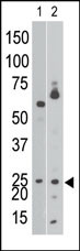
Applications |
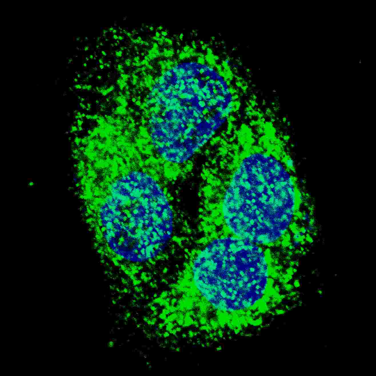
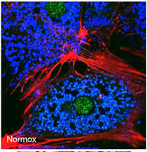
WB, IHC-P, IF, E
|
||
Dilution |
IF~~1:50~100
WB~~1:1000
IHC-P~~1:50~100
|
||
Synonyms |
BCL2/adenovirus E1B 19 kDa protein-interacting protein 3, BNIP3, NIP3
|
||
Images |
|
||
Specification |
|||
Quantity |
|
||
| Select | Brand | Catalog No. | Product Name | Pack Size | Type | Field of Research | Specification | Quantity | Price(USD) | |
| 1 | Leading Biology | APR03440G | ITGA11 Antibody (N-term) | 100 μl | Polyclonal Antibodies |
|
$495.00 | Add Ask | ||
| 2 | Leading Biology | APR04537G | CMIP Antibody (C-term) | 100 μl | Polyclonal Antibodies |
|
$495.00 | Add Ask | ||
| 3 | Leading Biology | APR12422G | Human H4 Histamine Receptor (extracellular) Antibody | 50 μl | Polyclonal Antibodies |
|
$695.00 | Add Ask | ||
| 4 | Leading Biology | APR03844G | UBE2W Antibody (C-term) | 100 μl | Polyclonal Antibodies |
|
$495.00 | Add Ask | ||
| 5 | Leading Biology | APR04349G | HECTD2 Antibody (N-term) | 100 μl | Polyclonal Antibodies |
|
$495.00 | Add Ask | ||
| 6 | Leading Biology | APR03502G | IGHG1 Antibody (Center) | 100 μl | Polyclonal Antibodies |
|
$495.00 | Add Ask |

You have 0 item in your cart
You have 0 item in your inquiry list