> Antigen, Antibodies, ELISA, Western Blot > Primary Antibody > Polyclonal Antibodies > BAI1 (extracellular) AntibodyBrand |
Leading Biology | Catalog Number |
APR00510G |
Product Type |
Polyclonal Antibodies | Field of Research |
|
Product Overview |
We constantly strive to ensure we provide our customers with the best antibodies. As a result of this work we offer this antibody in purified format.
We are in the process of updating our datasheets. If you have any questions regarding this update, please feel free to contact our technical support team.
This product is a high quality BAI1 (extracellular) Antibody.
|
||
Molecular Weight |
173297 Da
|
||
Host |
Rabbit
|
||
Species Reactivity |
Human, Mouse, Rat
|
||
Target |
Peptide (C)GRVRTYQFDSFLESTR, corresponding to amino acid residues 97-112 of mouse BAI1 (Accession Q3UHD1). Extracellular, N-terminus.The antibody will recognize the intact BAI1 receptor as well as the proteolytically processed N-terminal fragment. This fragment is also known as Vasculostatin (Vstat120).
|
||
GeneID |
|||
UniProt ID |
|||
Summary |
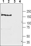
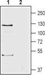
The three members of the brain angiogenesis inhibitor (BaI1-3) are receptors belonging to the adhesion subfamily of G-protein coupled receptor superfamily. Like all members of GPCRs, all three BaIs have seven transmembrane domains, an intracellular C-terminal tail and extracellular N-terminus. Like other adhesion members, the N-terminus is quite large1,2. Many domains are localized to the N-terminus; various glycosylations sites are present, there is a GPCR proteolysis site, a putative hormone binding domain and thrombospondin type 1 repeats which regulate the anti-angiogenic activity of thrombospondin-12,3. The C-terminal tail interacts with PDZ-domain proteins. Unique to BaI1 is a proline-rich domain required for interacting with Src homology domains and WW domain proteins2,4.Like most adhesion GPCRs, BaI also undergo proteolysis at the N-terminus at a highly rich cystein domain2. Following autocleavage, the N-terminal fragment remains associated to the receptor. In BaI1, proteolysis yields a partly secreted 120 kDa. fragment (vasculostatin-120) or a 40 kDa. fragment both having antiangiogenic effects2,5. At the mRNA level, all BaIs are expressed in fetal and adult human brain2,6. BaI2 is detected in the human heart and skeletal muscle. BaI3 is expressed in the human heart, testis and small intestine. In mouse, both BaI2 and BaI3 are restricted to the brain2.These receptors are implicated in various diseases and disorders such as primary glioma, pulmonary adenocarcinomas, gastric and colorectal cancers2,6,7.Abgent is pleased to offer a highly specific antibody directed against an extracellular epitope of mouse BaI1. Anti-Bai1 (extracellular) antibody (#AG1364) can be used in western blot and indirect flow cytometry applications. It has been designed to recognize Bai1 from rat, mouse, and human samples.
|
||
Form |
Liquid. Purified antibody supplied in 1x PBS buffer with 0.09% (w/v) sodium azide and 2% sucrose. |
||
Storage & Stability |
Store at +4°C short term. For long-term storage, aliquot and store at -20°C or below. Stable for 12 months at -20°C. Avoid repeated freeze-thaw cycles.
|
||
Applications |
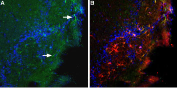
WB, IHC, FC
|
||
Dilution |
WB~~1:200-1:2000
IHC~~1:200
FC~~1:20
|
||
Images |
|
||
Specification |
|||
Quantity |
|
||
| Select | Brand | Catalog No. | Product Name | Pack Size | Type | Field of Research | Specification | Quantity | Price(USD) | |
| 1 | Leading Biology | APR03440G | ITGA11 Antibody (N-term) | 100 μl | Polyclonal Antibodies |
|
$495.00 | Add Ask | ||
| 2 | Leading Biology | APR04537G | CMIP Antibody (C-term) | 100 μl | Polyclonal Antibodies |
|
$495.00 | Add Ask | ||
| 3 | Leading Biology | APR12422G | Human H4 Histamine Receptor (extracellular) Antibody | 50 μl | Polyclonal Antibodies |
|
$695.00 | Add Ask | ||
| 4 | Leading Biology | APR03844G | UBE2W Antibody (C-term) | 100 μl | Polyclonal Antibodies |
|
$495.00 | Add Ask | ||
| 5 | Leading Biology | APR04349G | HECTD2 Antibody (N-term) | 100 μl | Polyclonal Antibodies |
|
$495.00 | Add Ask | ||
| 6 | Leading Biology | APR03502G | IGHG1 Antibody (Center) | 100 μl | Polyclonal Antibodies |
|
$495.00 | Add Ask |

You have 0 item in your cart
You have 0 item in your inquiry list