> Antigen, Antibodies, ELISA, Western Blot > Primary Antibody > Polyclonal Antibodies > VPAC1 (extracellular) AntibodyBrand |
Leading Biology | Catalog Number |
APR00487G |
Product Type |
Polyclonal Antibodies | Field of Research |
|
Product Overview |
We constantly strive to ensure we provide our customers with the best antibodies. As a result of this work we offer this antibody in purified format.
We are in the process of updating our datasheets. If you have any questions regarding this update, please feel free to contact our technical support team.
This product is a high quality VPAC1 (extracellular) Antibody.
|
||
Molecular Weight |
51547 Da
|
||
Host |
Rabbit
|
||
Species Reactivity |
Human, Mouse, Rat
|
||
Target |
Peptide (C)EEAQLENETIG(S)SK, corresponding to amino acid residues 52-65 of human VPAC1 (Accession P32241). Extracellular, N-terminus.
|
||
GeneID |
|||
UniProt ID |
|||
Summary |
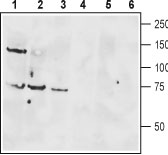
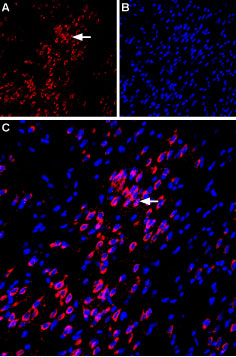
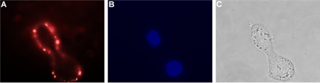
Vasointestinal peptide (VIP) and pituitary adenylate cyclase–activating peptide (PACAP) belong to the glucagon hormone superfamily, which includes secretin, growth hormone–releasing hormone (GHRH), glucagon, glucagon-like peptides 1 and 2 (GLP-1 and GLP-2), peptide histidine methionine (PHM), and glucose-dependent insulinotropic polypeptide (GIP)1. PACAP and VIP effects have been described in the digestive tract, cardiovascular system, airways, reproductive system, immune system, endocrine glands, and brain2. VIP and PACAP share a common G-protein coupled receptor, VPAC13.VPAC1 is a membrane-associated protein and shares significant homology with members of the G-protein coupled class B receptor family, the most important of which is the presence of large N-terminal extracellular domains which contain 10 highly conserved amino acids including six cysteines, putative N-terminal leader sequences and several potential N-glycosylation sites4. In the CNS, VPAC1 receptors are abundantly localized in piriform cortex, cerebral cortex, suprachiasmatic nucleus, hippocampus, and pineal gland5. In peripheral tissues, VPAC1 receptors have been found in breast, kidney, liver, lung, prostate, spleen, and mucosa of the gastrointestinal tract6. VPAC1 mediates a large array of VIP and PACAP actions on exocrine secretion, hormones release, muscle relaxation, metabolism, fetus growth, tumor cells and embryonic brain development7.Abgent is pleased to offer a highly specific antibody directed against an extracellular epitope of human VPAC1. Anti-VPAC1 (extracellular) antibody (#AG1002) can be used in western blot, immunohistochemistry,immunocytochemistry and indirect flow cytometry applications and has been designed to recognize VPAC1 from mouse, rat and human samples.
|
||
Form |
Liquid. Purified antibody supplied in 1x PBS buffer with 0.09% (w/v) sodium azide and 2% sucrose. |
||
Storage & Stability |
Store at +4°C short term. For long-term storage, aliquot and store at -20°C or below. Stable for 12 months at -20°C. Avoid repeated freeze-thaw cycles.
|
||
Applications |
WB, LCI
|
||
Dilution |
WB~~1:200-1:2000
IHC~~1:100
ICC~~1:25
FC~~1:20
|
||
Images |
|
||
Specification |
|||
Quantity |
|
||
| Select | Brand | Catalog No. | Product Name | Pack Size | Type | Field of Research | Specification | Quantity | Price(USD) | |
| 1 | Leading Biology | APR03440G | ITGA11 Antibody (N-term) | 100 μl | Polyclonal Antibodies |
|
$495.00 | Add Ask | ||
| 2 | Leading Biology | APR04537G | CMIP Antibody (C-term) | 100 μl | Polyclonal Antibodies |
|
$495.00 | Add Ask | ||
| 3 | Leading Biology | APR12422G | Human H4 Histamine Receptor (extracellular) Antibody | 50 μl | Polyclonal Antibodies |
|
$695.00 | Add Ask | ||
| 4 | Leading Biology | APR03844G | UBE2W Antibody (C-term) | 100 μl | Polyclonal Antibodies |
|
$495.00 | Add Ask | ||
| 5 | Leading Biology | APR04349G | HECTD2 Antibody (N-term) | 100 μl | Polyclonal Antibodies |
|
$495.00 | Add Ask | ||
| 6 | Leading Biology | APR03502G | IGHG1 Antibody (Center) | 100 μl | Polyclonal Antibodies |
|
$495.00 | Add Ask |

You have 0 item in your cart
You have 0 item in your inquiry list