> Antigen, Antibodies, ELISA, Western Blot > Primary Antibody > Polyclonal Antibodies > mGluR5 (extracellular) AntibodyBrand |
Leading Biology | Catalog Number |
APR00502G |
Product Type |
Polyclonal Antibodies | Field of Research |
|
Product Overview |
We constantly strive to ensure we provide our customers with the best antibodies. As a result of this work we offer this antibody in purified format.
We are in the process of updating our datasheets. If you have any questions regarding this update, please feel free to contact our technical support team.
This product is a high quality mGluR5 (extracellular) Antibody.
|
||
Molecular Weight |
131885 Da
|
||
Host |
Rabbit
|
||
Species Reactivity |
Mouse, Rat
|
||
Target |
Peptide EGFAQENSKYNKTC, corresponding to amino acids 367-380 of rat mGluR5 (Accession P31424). Extracellular, N-terminus.
|
||
GeneID |
|||
UniProt ID |
|||
Summary |
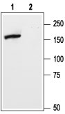
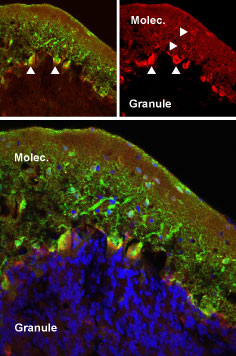
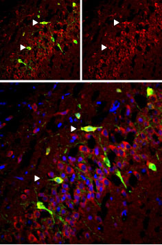
L-Glutamate, the major excitatory neurotransmitter in the central nervous system, operates through several receptors that are categorized as ionotropic (ligand-gated cation channels) or metabotropic (G-protein coupled receptors). The metabotropic glutamate receptors family includes eight members (mGluR1-8) that have been divided into three groups based on their sequence homology, pharmacology and signal transduction. Group I of the metabotropic glutamate receptors includes the mGluR1 and mGluR5 receptors. The receptors present the typical G-protein coupled receptor (GPCR) signature topology: seven transmembrane domains with a large extracellular N-terminus domain and an intracellular C-terminus one. The N-terminus domain of group I receptors contains the glutamate binding site while the cytoplasmic C-terminus domain has an important role in the regulation of receptor activity through interactions with other proteins such as the Homer adaptor proteins. mGluR1 and mGluR5 receptors signal through Gq/G11 that activates phospholipase C and ultimately produces an increase in inositol trisphosphate and cytosolic Ca2+. More downstream signaling pathways include activation of PKC and modulation of Ca2+ and K+ ion channels. Activation of signaling pathways independent of G-proteins has also been reported. mGluR5 is predominantly expressed in nervous tissue although expression in several non-neural cell types has also been described. In the brain it is highly expressed in the cortex, basal ganglia and hippocampus. The mGluR5 receptor is involved in several physiological processes such as neuronal development, induction of long-term potentiation (LTP) and depression (LTD) as well as in pathological disorders such as brain trauma, chronic pain, Parkinson’s and Huntington’s disease. Abgent is pleased to offer a highly specific antibody directed against the extracelluar N-terminus domain of the rat mGluR5 receptor. Anti-mGluR5 (extracellular) antibody (#AG1262) can be used in Western blot analysis, immunhistochemical and immunocytochemical applications, and recognizes mGluR5 in rat samples.
|
||
Form |
Liquid. Purified antibody supplied in 1x PBS buffer with 0.09% (w/v) sodium azide and 2% sucrose. |
||
Storage & Stability |
Store at +4°C short term. For long-term storage, aliquot and store at -20°C or below. Stable for 12 months at -20°C. Avoid repeated freeze-thaw cycles.
|
||
Applications |
WB, IHC, ICC, LCI
|
||
Dilution |
WB~~1:200-1:2000
IHC~~1:50
ICC~~1:100
|
||
Images |
|
||
Specification |
|||
Quantity |
|
||
| Select | Brand | Catalog No. | Product Name | Pack Size | Type | Field of Research | Specification | Quantity | Price(USD) | |
| 1 | Leading Biology | APR03440G | ITGA11 Antibody (N-term) | 100 μl | Polyclonal Antibodies |
|
$495.00 | Add Ask | ||
| 2 | Leading Biology | APR04537G | CMIP Antibody (C-term) | 100 μl | Polyclonal Antibodies |
|
$495.00 | Add Ask | ||
| 3 | Leading Biology | APR12422G | Human H4 Histamine Receptor (extracellular) Antibody | 50 μl | Polyclonal Antibodies |
|
$695.00 | Add Ask | ||
| 4 | Leading Biology | APR03844G | UBE2W Antibody (C-term) | 100 μl | Polyclonal Antibodies |
|
$495.00 | Add Ask | ||
| 5 | Leading Biology | APR04349G | HECTD2 Antibody (N-term) | 100 μl | Polyclonal Antibodies |
|
$495.00 | Add Ask | ||
| 6 | Leading Biology | APR03502G | IGHG1 Antibody (Center) | 100 μl | Polyclonal Antibodies |
|
$495.00 | Add Ask |

You have 0 item in your cart
You have 0 item in your inquiry list