> Antigen, Antibodies, ELISA, Western Blot > Primary Antibody > Polyclonal Antibodies > Goat Anti-ITCH / AIF4 AntibodyBrand |
Leading Biology | Catalog Number |
APG00174G |
Product Type |
Polyclonal Antibodies | Field of Research |
|
Product Overview |
We constantly strive to ensure we provide our customers with the best antibodies. As a result of this work we offer this antibody in purified format.
We are in the process of updating our datasheets. If you have any questions regarding this update, please feel free to contact our technical support team.
This product is a high quality Goat Anti-ITCH / AIF4 Antibody.
|
||
Molecular Weight |
102803 Da
|
||
Cellular Localization |
Antigen Cellular Localization:
Cell membrane. Cytoplasm. Nucleus. Note=Associates with endocytic vesicles. May be recruited to exosomes by NDFIP1
|
||
Host |
Goat
|
||
Species Reactivity |
Human
|
||
Isotype |
IgG
|
||
GeneID |
|||
UniProt ID |
|||
Function |
Acts as an E3 ubiquitin-protein ligase which accepts ubiquitin from an E2 ubiquitin-conjugating enzyme in the form of a thioester and then directly transfers the ubiquitin to targeted substrates. It catalyzes 'Lys-29'-, 'Lys-48'- and 'Lys-63'-linked ubiquitin conjugation. It is involved in the control of inflammatory signaling pathways. Is an essential component of a ubiquitin-editing protein complex, comprising also TNFAIP3, TAX1BP1 and RNF11, that ensures the transient nature of inflammatory signaling pathways. Promotes the association of the complex after TNF stimulation. Once the complex is formed, TNFAIP3 deubiquitinates 'Lys-63' polyubiquitin chains on RIPK1 and catalyzes the formation of 'Lys-48'-polyubiquitin chains. This leads to RIPK1 proteasomal degradation and consequently termination of the TNF- or LPS-mediated activation of NFKB1. Ubiquitinates RIPK2 by 'Lys-63'-linked conjugation and influences NOD2-dependent signal transduction pathways. Regulates the transcriptional activity of several transcription factors, and probably plays an important role in the regulation of immune response. Ubiquitinates NFE2 by 'Lys-63' linkages and is implicated in the control of the development of hematopoietic lineages. Critical regulator of T-helper (TH2) cytokine development through its ability to induce JUNB ubiquitination and degradation (By similarity). Ubiquitinates SNX9. Ubiquitinates CXCR4 and HGS/HRS and regulates sorting of CXCR4 to the degradative pathway. It is involved in the negative regulation of MAVS-dependent cellular antiviral responses. Ubiquitinates MAVS through 'Lys-48'-linked conjugation resulting in MAVS proteasomal degradation. Involved in the regulation of apoptosis and reactive oxygen species levels through the ubiquitination and proteasomal degradation of TXNIP. Mediates the antiapoptotic activity of epidermal growth factor through the ubiquitination and proteasomal degradation of p15 BID. Targets DTX1 for lysosomal degradation and controls NOTCH1 degradation, in the absence of ligand, through 'Lys-29'-linked polyubiquitination.
|
||
Summary |
Atrophin-1 contains a polyglutamine repeat, expansion of which is responsible for dentatorubral and pallidoluysian atrophy. The protein encoded by this gene interacts with atrophin-1. This encoded protein is a closely related member of the NEDD4-like protein family. This family of proteins are E3 ubiquitin-ligase molecules and regulate key trafficking decisions, including targeting of proteins to proteosomes or lysosomes. This encoded protein contains four tandem WW domains and a HECT (homologous to the E6-associated protein carboxyl terminus) domain. It can act as a transcriptional corepressor of p45/NFE2 and may participate in the regulation of immune responses by modifying Notch-mediated signaling. It is highly similar to the mouse Itch protein, which has been implicated in the regulation and differentiation of erythroid and lymphoid cells.
|
||
Form |
Liquid. Purified antibody supplied in 1x PBS buffer with 0.09% (w/v) sodium azide and 2% sucrose. |
||
Storage & Stability |
Store at +4°C short term. For long-term storage, aliquot and store at -20°C or below. Stable for 12 months at -20°C. Avoid repeated freeze-thaw cycles.
|
||
Applications |
WB, E
|
||
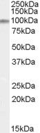
Images |
|
||
Specification |
|||
Quantity |
|
||
| Select | Brand | Catalog No. | Product Name | Pack Size | Type | Field of Research | Specification | Quantity | Price(USD) | |
| 1 | Leading Biology | APR03440G | ITGA11 Antibody (N-term) | 100 μl | Polyclonal Antibodies |
|
$495.00 | Add Ask | ||
| 2 | Leading Biology | APR04537G | CMIP Antibody (C-term) | 100 μl | Polyclonal Antibodies |
|
$495.00 | Add Ask | ||
| 3 | Leading Biology | APR12422G | Human H4 Histamine Receptor (extracellular) Antibody | 50 μl | Polyclonal Antibodies |
|
$695.00 | Add Ask | ||
| 4 | Leading Biology | APR03844G | UBE2W Antibody (C-term) | 100 μl | Polyclonal Antibodies |
|
$495.00 | Add Ask | ||
| 5 | Leading Biology | APR04349G | HECTD2 Antibody (N-term) | 100 μl | Polyclonal Antibodies |
|
$495.00 | Add Ask | ||
| 6 | Leading Biology | APR03502G | IGHG1 Antibody (Center) | 100 μl | Polyclonal Antibodies |
|
$495.00 | Add Ask |

You have 0 item in your cart
You have 0 item in your inquiry list