> Antigen, Antibodies, ELISA, Western Blot > Primary Antibody > Polyclonal Antibodies > Goat Anti-FGR AntibodyBrand |
Leading Biology | Catalog Number |
APG00124G |
Product Type |
Polyclonal Antibodies | Field of Research |
|
Product Overview |
We constantly strive to ensure we provide our customers with the best antibodies. As a result of this work we offer this antibody in purified format.
We are in the process of updating our datasheets. If you have any questions regarding this update, please feel free to contact our technical support team.
This product is a high quality Goat Anti-FGR Antibody.
|
||
Molecular Weight |
59479 Da
|
||
Cellular Localization |
Antigen Cellular Localization:
Cell membrane; Lipid-anchor; Cytoplasmic side. Cell membrane; Peripheral membrane protein; Cytoplasmic side. Cell projection, ruffle membrane. Cytoplasm, cytosol. Cytoplasm, cytoskeleton Mitochondrion inner membrane. Mitochondrion intermembrane space. Note=Detected in mitochondrial intermembrane space and at inner membranes (By similarity) Colocalizes with actin fibers at membrane ruffles. Detected at plasma membrane lipid rafts.
|
||
Host |
Goat
|
||
Species Reactivity |
Mouse
|
||
Isotype |
IgG
|
||
Symbol |
SRC2
|
||
GeneID |
|||
UniProt ID |
|||
Function |
Non-receptor tyrosine-protein kinase that transmits signals from cell surface receptors devoid of kinase activity and contributes to the regulation of immune responses, including neutrophil, monocyte, macrophage and mast cell functions, cytoskeleton remodeling in response to extracellular stimuli, phagocytosis, cell adhesion and migration. Promotes mast cell degranulation, release of inflammatory cytokines and IgE-mediated anaphylaxis. Acts downstream of receptors that bind the Fc region of immunoglobulins, such as MS4A2/FCER1B, FCGR2A and/or FCGR2B. Acts downstream of ITGB1 and ITGB2, and regulates actin cytoskeleton reorganization, cell spreading and adhesion. Depending on the context, activates or inhibits cellular responses. Functions as negative regulator of ITGB2 signaling, phagocytosis and SYK activity in monocytes. Required for normal ITGB1 and ITGB2 signaling, normal cell spreading and adhesion in neutrophils and macrophages. Functions as positive regulator of cell migration and regulates cytoskeleton reorganization via RAC1 activation. Phosphorylates SYK (in vitro) and promotes SYK- dependent activation of AKT1 and MAP kinase signaling. Phosphorylates PLD2 in antigen-stimulated mast cells, leading to PLD2 activation and the production of the signaling molecules lysophosphatidic acid and diacylglycerol. Promotes activation of PIK3R1. Phosphorylates FASLG, and thereby regulates its ubiquitination and subsequent internalization. Phosphorylates ABL1. Promotes phosphorylation of CBL, CTTN, PIK3R1, PTK2/FAK1, PTK2B/PYK2 and VAV2. Phosphorylates HCLS1 that has already been phosphorylated by SYK, but not unphosphorylated HCLS1.
|
||
Summary |
This gene is a member of the Src family of protein tyrosine kinases (PTKs). The encoded protein contains N-terminal sites for myristylation and palmitylation, a PTK domain, and SH2 and SH3 domains which are involved in mediating protein-protein interactions with phosphotyrosine-containing and proline-rich motifs, respectively. The protein localizes to plasma membrane ruffles, and functions as a negative regulator of cell migration and adhesion triggered by the beta-2 integrin signal transduction pathway. Infection with Epstein-Barr virus results in the overexpression of this gene. Multiple alternatively spliced variants, encoding the same protein, have been identified.
|
||
Form |
Liquid. Purified antibody supplied in 1x PBS buffer with 0.09% (w/v) sodium azide and 2% sucrose. |
||
Storage & Stability |
Store at +4°C short term. For long-term storage, aliquot and store at -20°C or below. Stable for 12 months at -20°C. Avoid repeated freeze-thaw cycles.
|
||
Applications |
WB, E
|
||
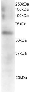
Images |
|
||
Specification |
|||
Quantity |
|
||
| Select | Brand | Catalog No. | Product Name | Pack Size | Type | Field of Research | Specification | Quantity | Price(USD) | |
| 1 | Leading Biology | APR03440G | ITGA11 Antibody (N-term) | 100 μl | Polyclonal Antibodies |
|
$495.00 | Add Ask | ||
| 2 | Leading Biology | APR04537G | CMIP Antibody (C-term) | 100 μl | Polyclonal Antibodies |
|
$495.00 | Add Ask | ||
| 3 | Leading Biology | APR12422G | Human H4 Histamine Receptor (extracellular) Antibody | 50 μl | Polyclonal Antibodies |
|
$695.00 | Add Ask | ||
| 4 | Leading Biology | APR03844G | UBE2W Antibody (C-term) | 100 μl | Polyclonal Antibodies |
|
$495.00 | Add Ask | ||
| 5 | Leading Biology | APR04349G | HECTD2 Antibody (N-term) | 100 μl | Polyclonal Antibodies |
|
$495.00 | Add Ask | ||
| 6 | Leading Biology | APR03502G | IGHG1 Antibody (Center) | 100 μl | Polyclonal Antibodies |
|
$495.00 | Add Ask |

You have 0 item in your cart
You have 0 item in your inquiry list