> Antigen, Antibodies, ELISA, Western Blot > Primary Antibody > Polyclonal Antibodies > Goat Anti-Cortactin / EMS1 AntibodyBrand |
Leading Biology | Catalog Number |
APG00079G |
Product Type |
Polyclonal Antibodies | Field of Research |
|
Product Overview |
We constantly strive to ensure we provide our customers with the best antibodies. As a result of this work we offer this antibody in purified format.
We are in the process of updating our datasheets. If you have any questions regarding this update, please feel free to contact our technical support team.
This product is a high quality Goat Anti-Cortactin / EMS1 Antibody.
|
||
Molecular Weight |
61586 Da
|
||
Cellular Localization |
Antigen Cellular Localization:
Cytoplasm, cytoskeleton. Cell projection, lamellipodium. Cell projection, ruffle. Cell projection, dendrite. Cell projection {ECO:0000250|UniProtKB:Q66HL2}. Cell membrane; Peripheral membrane protein; Cytoplasmic side. Cell projection, podosome {ECO:0000250|UniProtKB:Q01406}. Cell junction {ECO:0000250|UniProtKB:Q66HL2}. Cell junction, focal adhesion {ECO:0000250|UniProtKB:Q66HL2}. Membrane, clathrin-coated pit {ECO:0000250|UniProtKB:Q66HL2}. Cell projection, dendritic spine. Cytoplasm, cell cortex. Note=Colocalizes transiently with PTK2/FAK1 at focal adhesions (By similarity). Associated with membrane ruffles and lamellipodia. In the presence of CTTNBP2NL, colocalizes with stress fibers (By similarity). In the presence of CTTNBP2, localizes at the cell cortex (By similarity). In response to neuronal activation by glutamate, redistributes from dendritic spines to the dendritic shaft (By similarity). Colocalizes with DNM2 at the basis of filopodia in hippocampus neuron growth zones (By similarity). {ECO:0000250|UniProtKB:Q60598, ECO:0000250|UniProtKB:Q66HL2}
|
||
Host |
Goat
|
||
Species Reactivity |
Mouse
|
||
Isotype |
IgG
|
||
Symbol |
EMS1
|
||
GeneID |
|||
UniProt ID |
|||
Function |
Contributes to the organization of the actin cytoskeleton and cell shape (PubMed:21296879). Plays a role in the formation of lamellipodia and in cell migration. Plays a role in the regulation of neuron morphology, axon growth and formation of neuronal growth cones (By similarity). Through its interaction with CTTNBP2, involved in the regulation of neuronal spine density (By similarity). Plays a role in the invasiveness of cancer cells, and the formation of metastases (PubMed:16636290). Plays a role in focal adhesion assembly and turnover (By similarity). In complex with ABL1 and MYLK regulates cortical actin-based cytoskeletal rearrangement critical to sphingosine 1-phosphate (S1P)-mediated endothelial cell (EC) barrier enhancement (PubMed:20861316). Plays a role in intracellular protein transport and endocytosis, and in modulating the levels of potassium channels present at the cell membrane (PubMed:17959782). Plays a role in receptor-mediated endocytosis via clathrin-coated pits (By similarity).
|
||
Summary |
This gene is overexpressed in breast cancer and squamous cell carcinomas of the head and neck. The encoded protein is localized in the cytoplasm and in areas of the cell-substratum contacts. This gene has two roles: (1) regulating the interactions between components of adherens-type junctions and (2) organizing the cytoskeleton and cell adhesion structures of epithelia and carcinoma cells. During apoptosis, the encoded protein is degraded in a caspase-dependent manner. The aberrant regulation of this gene contributes to tumor cell invasion and metastasis. Three splice variants that encode different isoforms have been identified for this gene.
|
||
Form |
Liquid. Purified antibody supplied in 1x PBS buffer with 0.09% (w/v) sodium azide and 2% sucrose. |
||
Storage & Stability |
Store at +4°C short term. For long-term storage, aliquot and store at -20°C or below. Stable for 12 months at -20°C. Avoid repeated freeze-thaw cycles.
|
||
Applications |
WB, E
|
||
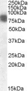
Images |
|
||
Specification |
|||
Quantity |
|
||
| Select | Brand | Catalog No. | Product Name | Pack Size | Type | Field of Research | Specification | Quantity | Price(USD) | |
| 1 | Leading Biology | APR03440G | ITGA11 Antibody (N-term) | 100 μl | Polyclonal Antibodies |
|
$495.00 | Add Ask | ||
| 2 | Leading Biology | APR04537G | CMIP Antibody (C-term) | 100 μl | Polyclonal Antibodies |
|
$495.00 | Add Ask | ||
| 3 | Leading Biology | APR12422G | Human H4 Histamine Receptor (extracellular) Antibody | 50 μl | Polyclonal Antibodies |
|
$695.00 | Add Ask | ||
| 4 | Leading Biology | APR03844G | UBE2W Antibody (C-term) | 100 μl | Polyclonal Antibodies |
|
$495.00 | Add Ask | ||
| 5 | Leading Biology | APR04349G | HECTD2 Antibody (N-term) | 100 μl | Polyclonal Antibodies |
|
$495.00 | Add Ask | ||
| 6 | Leading Biology | APR03502G | IGHG1 Antibody (Center) | 100 μl | Polyclonal Antibodies |
|
$495.00 | Add Ask |

You have 0 item in your cart
You have 0 item in your inquiry list