> Antigen, Antibodies, ELISA, Western Blot > Primary Antibody > Polyclonal Antibodies > MST1 Antibody (C-term)Brand |
Leading Biology | Catalog Number |
APR06032G |
Product Type |
Polyclonal Antibodies | Field of Research |
|
Product Overview |
We constantly strive to ensure we provide our customers with the best antibodies. As a result of this work we offer this antibody in purified format.
We are in the process of updating our datasheets. If you have any questions regarding this update, please feel free to contact our technical support team.
This product is a high quality MST1 Antibody (C-term).
|
||
Molecular Weight |
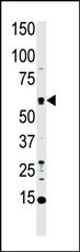
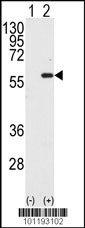
55630 Da
|
||
Cellular Localization |
Antigen Cellular Localization:
Cytoplasm. Nucleus. Note=The caspase-cleaved form cycles between the nucleus and cytoplasm
|
||
Host |
Rabbit
|
||
Species Reactivity |
Human
|
||
Immunogen |
385-415 aa
|
||
Target |
This MST1 antibody is generated from rabbits immunized with a KLH conjugated synthetic peptide between 385-415 amino acids from the C-terminal region of human MST1.
|
||
Isotype |
Rabbit Ig
|
||
Symbol |
KRS2, MST1
|
||
GeneID |
|||
UniProt ID |
|||
Function |
Stress-activated, pro-apoptotic kinase which, following caspase-cleavage, enters the nucleus and induces chromatin condensation followed by internucleosomal DNA fragmentation. Key component of the Hippo signaling pathway which plays a pivotal role in organ size control and tumor suppression by restricting proliferation and promoting apoptosis. The core of this pathway is composed of a kinase cascade wherein STK3/MST2 and STK4/MST1, in complex with its regulatory protein SAV1, phosphorylates and activates LATS1/2 in complex with its regulatory protein MOB1, which in turn phosphorylates and inactivates YAP1 oncoprotein and WWTR1/TAZ. Phosphorylation of YAP1 by LATS2 inhibits its translocation into the nucleus to regulate cellular genes important for cell proliferation, cell death, and cell migration. STK3/MST2 and STK4/MST1 are required to repress proliferation of mature hepatocytes, to prevent activation of facultative adult liver stem cells (oval cells), and to inhibit tumor formation (By similarity). Phosphorylates 'Ser-14' of histone H2B (H2BS14ph) during apoptosis. Phosphorylates FOXO3 upon oxidative stress, which results in its nuclear translocation and cell death initiation. Phosphorylates MOBKL1A, MOBKL1B and RASSF2. Phosphorylates TNNI3 (cardiac Tn-I) and alters its binding affinity to TNNC1 (cardiac Tn-C) and TNNT2 (cardiac Tn-T). Phosphorylates FOXO1 on 'Ser-212' and regulates its activation and stimulates transcription of PMAIP1 in a FOXO1-dependent manner. Phosphorylates SIRT1 and inhibits SIRT1-mediated p53/TP53 deacetylation, thereby promoting p53/TP53 dependent transcription and apoptosis upon DNA damage. Acts as an inhibitor of PKB/AKT1. Phosphorylates AR on 'Ser-650' and suppresses its activity by intersecting with PKB/AKT1 signaling and antagonizing formation of AR-chromatin complexes.
|
||
Summary |
MST1 is a cytoplasmic kinase that is structurally similar to the yeast Ste20p kinase, which acts upstream of the stress-induced mitogen-activated protein kinase cascade. The encoded protein can phosphorylate myelin basic protein and undergoes autophosphorylation. A caspase-cleaved fragment of the encoded protein has been shown to be capable of phosphorylating histone H2B. The particular phosphorylation catalyzed by this protein has been correlated with apoptosis, and it is possible that this protein induces the chromatin condensation observed in this process.
|
||
Storage & Stability |
Store at +4°C short term. For long-term storage, aliquot and store at -20°C or below. Stable for 12 months at -20°C. Avoid repeated freeze-thaw cycles.
|
||
Applications |
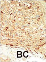
WB, IHC-P, E
|
||
Dilution |
WB~~1:1000
IHC-P~~1:50~100
|
||
Images |
|
||
Specification |
|||
Quantity |
|
||
| Select | Brand | Catalog No. | Product Name | Pack Size | Type | Field of Research | Specification | Quantity | Price(USD) | |
| 1 | Leading Biology | APR03440G | ITGA11 Antibody (N-term) | 100 μl | Polyclonal Antibodies |
|
$495.00 | Add Ask | ||
| 2 | Leading Biology | APR04537G | CMIP Antibody (C-term) | 100 μl | Polyclonal Antibodies |
|
$495.00 | Add Ask | ||
| 3 | Leading Biology | APR12422G | Human H4 Histamine Receptor (extracellular) Antibody | 50 μl | Polyclonal Antibodies |
|
$695.00 | Add Ask | ||
| 4 | Leading Biology | APR03844G | UBE2W Antibody (C-term) | 100 μl | Polyclonal Antibodies |
|
$495.00 | Add Ask | ||
| 5 | Leading Biology | APR04349G | HECTD2 Antibody (N-term) | 100 μl | Polyclonal Antibodies |
|
$495.00 | Add Ask | ||
| 6 | Leading Biology | APR03502G | IGHG1 Antibody (Center) | 100 μl | Polyclonal Antibodies |
|
$495.00 | Add Ask |

You have 0 item in your cart
You have 0 item in your inquiry list