> Antigen, Antibodies, ELISA, Western Blot > Primary Antibody > Polyclonal Antibodies > CXCR3 Antibody (Center)Brand |
Leading Biology | Catalog Number |
APR03422G |
Product Type |
Polyclonal Antibodies | Field of Research |
|
Product Overview |
We constantly strive to ensure we provide our customers with the best antibodies. As a result of this work we offer this antibody in purified format.
We are in the process of updating our datasheets. If you have any questions regarding this update, please feel free to contact our technical support team.
This product is a high quality CXCR3 Antibody (Center).
|
||
Molecular Weight |
40660 Da
|
||
Cellular Localization |
Antigen Cellular Localization:
Isoform 1: Cell membrane; Multi-pass membrane protein
|
||
Host |
Rabbit
|
||
Species Reactivity |
Human
|
||
Immunogen |
140-167 aa
|
||
Target |
This CXCR3 antibody is generated from rabbits immunized with a KLH conjugated synthetic peptide between 140-167 amino acids from the Central region of human CXCR3.
|
||
Isotype |
Rabbit Ig
|
||
Symbol |
GPR9
|
||
GeneID |
|||
UniProt ID |
|||
Function |
Isoform 1: Receptor for the C-X-C chemokine CXCL9, CXCL10 and CXCL11 and mediates the proliferation, survival and angiogenic activity of human mesangial cells (HMC) through a heterotrimeric G-protein signaling pathway (PubMed:12782716). Binds to CCL21. Probably promotes cell chemotaxis response. Isoform 3: Mediates the activity of CXCL11.
|
||
Summary |
This gene encodes a G protein-coupled receptor withselectivity for three chemokines, termed IP10(interferon-g-inducible 10 kDa protein), Mig (monokine induced byinterferon-g) and I-TAC (interferon-inducible T cella-chemoattractant). IP10, Mig and I-TAC belong to the structuralsubfamily of CXC chemokines, in which a single amino acid residueseparates the first two of four highly conserved Cys residues.Binding of chemokines to this protein induces cellular responsesthat are involved in leukocyte traffic, most notably integrinactivation, cytoskeletal changes and chemotactic migration.Inhibition by Bordetella pertussis toxin suggests thatheterotrimeric G protein of the Gi-subclass couple to this protein.Signal transduction has not been further analyzed but may includethe same enzymes that were identified in the signaling cascadeinduced by other chemokine receptors. As a consequence ofchemokine-induced cellular desensitization(phosphorylation-dependent receptor internalization), cellularresponses are typically rapid and short in duration. Cellularresponsiveness is restored after dephosphorylation of intracellularreceptors and subsequent recycling to the cell surface. This geneis prominently expressed in in vitro cultured effector/memory Tcells, and in T cells present in many types of inflamed tissues. Inaddition, IP10, Mig and I-TAC are commonly produced by local cellsin inflammatory lesion, suggesting that this gene and itschemokines participate in the recruitment of inflammatory cells.Therefore, this protein is a target for the development of smallmolecular weight antagonists, which may be used in the treatment ofdiverse inflammatory diseases. Multiple transcript variantsencoding different isoforms have been found for this gene.
|
||
Storage & Stability |
Store at +4°C short term. For long-term storage, aliquot and store at -20°C or below. Stable for 12 months at -20°C. Avoid repeated freeze-thaw cycles.
|
||
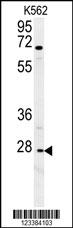
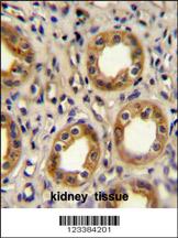
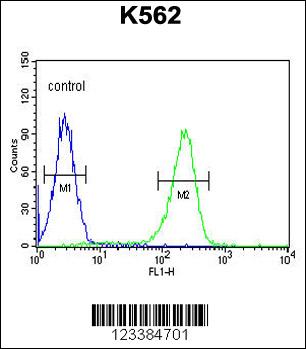
Applications |
WB, IHC-P, FC, E
|
||
Dilution |
WB~~1:1000
IHC-P~~1:50~100
FC~~1:10~50
|
||
Images |
|
||
Specification |
|||
Quantity |
|
||
| Select | Brand | Catalog No. | Product Name | Pack Size | Type | Field of Research | Specification | Quantity | Price(USD) | |
| 1 | Leading Biology | APR03440G | ITGA11 Antibody (N-term) | 100 μl | Polyclonal Antibodies |
|
$495.00 | Add Ask | ||
| 2 | Leading Biology | APR04537G | CMIP Antibody (C-term) | 100 μl | Polyclonal Antibodies |
|
$495.00 | Add Ask | ||
| 3 | Leading Biology | APR12422G | Human H4 Histamine Receptor (extracellular) Antibody | 50 μl | Polyclonal Antibodies |
|
$695.00 | Add Ask | ||
| 4 | Leading Biology | APR03844G | UBE2W Antibody (C-term) | 100 μl | Polyclonal Antibodies |
|
$495.00 | Add Ask | ||
| 5 | Leading Biology | APR04349G | HECTD2 Antibody (N-term) | 100 μl | Polyclonal Antibodies |
|
$495.00 | Add Ask | ||
| 6 | Leading Biology | APR03502G | IGHG1 Antibody (Center) | 100 μl | Polyclonal Antibodies |
|
$495.00 | Add Ask |

You have 0 item in your cart
You have 0 item in your inquiry list