> Antigen, Antibodies, ELISA, Western Blot > Primary Antibody > Monoclonal Antibodies > MCL-1 AntibodyBrand |
Leading Biology | Catalog Number |
APR08381G |
Product Type |
Monoclonal Antibodies | Field of Research |
|
Product Overview |
We constantly strive to ensure we provide our customers with the best antibodies. As a result of this work we offer this antibody in purified format.
We are in the process of updating our datasheets. If you have any questions regarding this update, please feel free to contact our technical support team.
This product is a high quality MCL-1 antibody.
|
||
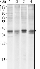
Molecular Weight |
37kDa
|
||
Cellular Localization |
Antigen Cellular Localization:
Membrane; Single-pass membrane protein. Cytoplasm. Mitochondrion. Nucleus, nucleoplasm. Note=Cytoplasmic, associated with mitochondria
|
||
Host |
Mouse
|
||
Species Reactivity |
Human
|
||
Clone |
8C6; 8C6D4B1
|
||
Isotype |
IgG1
|
||
Symbol |
BCL2L3
|
||
GeneID |
|||
UniProt ID |
|||
Function |
Involved in the regulation of apoptosis versus cell survival, and in the maintenance of viability but not of proliferation. Mediates its effects by interactions with a number of other regulators of apoptosis. Isoform 1 inhibits apoptosis. Isoform 2 promotes apoptosis.
|
||
Form |
Liquid. In PBS containing 0.02% sodium azide. |
||
Storage & Stability |
Store at +4°C short term. For long-term storage, aliquot and store at -20°C or below. Stable for 12 months at -20°C. Avoid repeated freeze-thaw cycles.
|
||
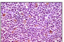
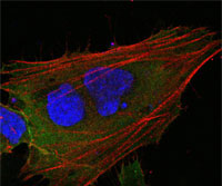
Applications |
WB, IHC, ICC, E
|
||
Dilution |
WB~~1/500 - 1/2000
IHC~~1/200 - 1/1000
IF~~1/200 - 1/1000
|
||
Synonyms |
Induced myeloid leukemia cell differentiation protein Mcl-1, Bcl-2-like protein 3, Bcl2-L-3, Bcl-2-related protein EAT/mcl1, mcl1/EAT, MCL1, BCL2L3
|
||
Images |
|
||
Specification |
|||
Quantity |
|
||
| Select | Brand | Catalog No. | Product Name | Pack Size | Type | Field of Research | Specification | Quantity | Price(USD) | |
| 1 | Leading Biology | APG02467G | CCK4 / PTK7 Antibody (clone 4F9) | 50 μl | Monoclonal Antibodies |
|
$495.00 | Add Ask | ||
| 2 | Leading Biology | AMM04683G | GALT Antibody (clone 4C11) | 50 μg | Monoclonal Antibodies |
|
$545.00 | Add Ask | ||
| 3 | Leading Biology | AMM01402G | Vimentin (Mesenchymal Cell Marker) Antibody - With BSA and Azide | 50 ug | Monoclonal Antibodies |
|
$395.00 | Add Ask | ||
| 4 | Leading Biology | APR08280G | LTA4H / LTA4 Antibody (clone 9G8) | 50 μl | Monoclonal Antibodies |
|
$495.00 | Add Ask | ||
| 5 | Leading Biology | AMM00172G | CD1a / HTA1 (Mature Langerhans Cells Marker) Antibody - With BSA and Azide | 50 ug | Monoclonal Antibodies |
|
$395.00 | Add Ask | ||
| 6 | Leading Biology | AMM05750G | CEBPA Antibody | 100 μl | Monoclonal Antibodies |
|
$545.00 | Add Ask |

You have 0 item in your cart
You have 0 item in your inquiry list