> Antigen, Antibodies, ELISA, Western Blot > Primary Antibody > Monoclonal Antibodies > MYOC / Myocilin Antibody (clone 4F8)Brand |
Leading Biology | Catalog Number |
AMM06528G |
Product Type |
Monoclonal Antibodies | Field of Research |
|
Product Overview |
We constantly strive to ensure we provide our customers with the best antibodies. As a result of this work we offer this antibody in purified format.
We are in the process of updating our datasheets. If you have any questions regarding this update, please feel free to contact our technical support team.
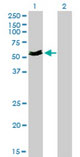
This product is a high quality MYOC / Myocilin antibody (clone 4F8).
|
||
Molecular Weight |
57kDa
|
||
Cellular Localization |
Antigen Cellular Localization:
Secreted. Golgi apparatus. Cytoplasmic vesicle. Secreted, extracellular space. Secreted, extracellular space, extracellular matrix. Secreted, exosome. Mitochondrion Mitochondrion intermembrane space. Mitochondrion inner membrane Mitochondrion outer membrane. Rough endoplasmic reticulum. Cell projection. Cell projection, cilium. Note=Located preferentially in the ciliary rootlet and basal body of the connecting cilium of photoreceptor cells, and in the rough endoplasmic reticulum. It is only imported to mitochondria in the trabecular meshwork Localizes to the Golgi apparatus in Schlemm's canal endothelial cells. Appears in the extracellular space of trabecular meshwork cells by an unconventional mechanism, likely associated with exosome-like vesicles. Localizes in trabecular meshwork extracellular matrix Myocilin, N-terminal fragment: Endoplasmic reticulum. Note=Remains retained in the endoplasmic reticulum
|
||
Host |
Mouse
|
||
Species Reactivity |
Human
|
||
Clone |
4F8
|
||
Symbol |
GLC1A, TIGR
|
||
GeneID |
|||
UniProt ID |
|||
Function |
Secreted glycoprotein regulating the activation of different signaling pathways in adjacent cells to control different processes including cell adhesion, cell-matrix adhesion, cytoskeleton organization and cell migration. Promotes substrate adhesion, spreading and formation of focal contacts. Negatively regulates cell-matrix adhesion and stress fiber assembly through Rho protein signal transduction. Modulates the organization of actin cytoskeleton by stimulating the formation of stress fibers through interactions with components of Wnt signaling pathways. Promotes cell migration through activation of PTK2 and the downstream phosphatidylinositol 3-kinase signaling. Plays a role in bone formation and promotes osteoblast differentiation in a dose-dependent manner through mitogen-activated protein kinase signaling. Mediates myelination in the peripheral nervous system through ERBB2/ERBB3 signaling. Plays a role as a regulator of muscle hypertrophy through the components of dystrophin-associated protein complex. Involved in positive regulation of mitochondrial depolarization. Plays a role in neurite outgrowth. May participate in the obstruction of fluid outflow in the trabecular meshwork.
|
||
Summary |
Secreted glycoprotein regulating the activation of different signaling pathways in adjacent cells to control different processes including cell adhesion, cell-matrix adhesion, cytoskeleton organization and cell migration. Promotes substrate adhesion, spreading and formation of focal contacts. Negatively regulates cell-matrix adhesion and stress fiber assembly through Rho protein signal transduction. Modulates the organization of actin cytoskeleton by stimulating the formation of stress fibers through interactions with components of Wnt signaling pathways. Promotes cell migration through activation of PTK2 and the downstream phosphatidylinositol 3-kinase signaling. Plays a role in bone formation and promotes osteoblast differentiation in a dose-dependent manner through mitogen-activated protein kinase signaling. Mediates myelination in the peripheral nervous system through ERBB2/ERBB3 signaling. Plays a role as a regulator of muscle hypertrophy through the components of dystrophin-associated protein complex. Involved in positive regulation of mitochondrial depolarization. Plays a role in neurite outgrowth. May participate in the obstruction of fluid outflow in the trabecular meshwork.
|
||
Form |
Liquid |
||
Storage & Stability |
Store at +4°C short term. For long-term storage, aliquot and store at -20°C or below. Stable for 12 months at -20°C. Avoid repeated freeze-thaw cycles.
|
||
Applications |
WB, IHC-P, E, RNAi
|
||
Dilution |
IHC-P (5 μg/ml)
|
||
Synonyms |
Myocilin, Myocilin 55 kDa subunit, Trabecular meshwork-induced glucocorticoid response protein, Myocilin, N-terminal fragment, Myocilin 20 kDa N-terminal fragment, Myocilin, C-terminal fragment, Myocilin 35 kDa N-terminal fragment, MYOC, GLC1A, TIGR
|
||
Images |
|
||
Specification |
|||
Quantity |
|
||
| Select | Brand | Catalog No. | Product Name | Pack Size | Type | Field of Research | Specification | Quantity | Price(USD) | |
| 1 | Leading Biology | APG02467G | CCK4 / PTK7 Antibody (clone 4F9) | 50 μl | Monoclonal Antibodies |
|
$495.00 | Add Ask | ||
| 2 | Leading Biology | AMM04683G | GALT Antibody (clone 4C11) | 50 μg | Monoclonal Antibodies |
|
$545.00 | Add Ask | ||
| 3 | Leading Biology | AMM01402G | Vimentin (Mesenchymal Cell Marker) Antibody - With BSA and Azide | 50 ug | Monoclonal Antibodies |
|
$395.00 | Add Ask | ||
| 4 | Leading Biology | APR08280G | LTA4H / LTA4 Antibody (clone 9G8) | 50 μl | Monoclonal Antibodies |
|
$495.00 | Add Ask | ||
| 5 | Leading Biology | AMM00172G | CD1a / HTA1 (Mature Langerhans Cells Marker) Antibody - With BSA and Azide | 50 ug | Monoclonal Antibodies |
|
$395.00 | Add Ask | ||
| 6 | Leading Biology | AMM05750G | CEBPA Antibody | 100 μl | Monoclonal Antibodies |
|
$545.00 | Add Ask |

You have 0 item in your cart
You have 0 item in your inquiry list