> Antigen, Antibodies, ELISA, Western Blot > Primary Antibody > Monoclonal Antibodies > CDK9 AntibodyBrand |
Leading Biology | Catalog Number |
APG02543G |
Product Type |
Monoclonal Antibodies | Field of Research |
|
Product Overview |
We constantly strive to ensure we provide our customers with the best antibodies. As a result of this work we offer this antibody in purified format.
We are in the process of updating our datasheets. If you have any questions regarding this update, please feel free to contact our technical support team.
This product is a high quality CDK9 antibody.
|
||
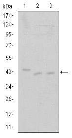
Molecular Weight |
43kDa
|
||
Cellular Localization |
Antigen Cellular Localization:
Nucleus. Cytoplasm. Nucleus, PML body. Note=Accumulates on chromatin in response to replication stress Complexed with CCNT1 in nuclear speckles, but uncomplexed form in the cytoplasm. The translocation from nucleus to cytoplasm is XPO1/CRM1-dependent. Associates with PML body when acetylated
|
||
Host |
Mouse
|
||
Species Reactivity |
Human
|
||
Clone |
1B5D10
|
||
Isotype |
IgG1
|
||
Symbol |
CDC2L4, TAK
|
||
GeneID |
|||
UniProt ID |
|||
Function |
Protein kinase involved in the regulation of transcription. Member of the cyclin-dependent kinase pair (CDK9/cyclin-T) complex, also called positive transcription elongation factor b (P-TEFb), which facilitates the transition from abortive to productive elongation by phosphorylating the CTD (C-terminal domain) of the large subunit of RNA polymerase II (RNAP II) POLR2A, SUPT5H and RDBP. This complex is inactive when in the 7SK snRNP complex form. Phosphorylates EP300, MYOD1, RPB1/POLR2A and AR, and the negative elongation factors DSIF and NELF. Regulates cytokine inducible transcription networks by facilitating promoter recognition of target transcription factors (e.g. TNF-inducible RELA/p65 activation and IL-6-inducible STAT3 signaling). Promotes RNA synthesis in genetic programs for cell growth, differentiation and viral pathogenesis. P-TEFb is also involved in cotranscriptional histone modification, mRNA processing and mRNA export. Modulates a complex network of chromatin modifications including histone H2B monoubiquitination (H2Bub1), H3 lysine 4 trimethylation (H3K4me3) and H3K36me3; integrates phosphorylation during transcription with chromatin modifications to control co-transcriptional histone mRNA processing. The CDK9/cyclin-K complex has also a kinase activity towards CTD of RNAP II and can substitute for CDK9/cyclin-T P-TEFb in vitro. Replication stress response protein; the CDK9/cyclin-K complex is required for genome integrity maintenance, by promoting cell cycle recovery from replication arrest and limiting single- stranded DNA amount in response to replication stress, thus reducing the breakdown of stalled replication forks and avoiding DNA damage. In addition, probable function in DNA repair of isoform 2 via interaction with KU70/XRCC6. Promotes cardiac myocyte enlargement. RPB1/POLR2A phosphorylation on 'Ser-2' in CTD activates transcription. AR phosphorylation modulates AR transcription factor promoter selectivity and cell growth. DSIF and NELF phosphorylation promotes transcription by inhibiting their negative effect. The phosphorylation of MYOD1 enhances its transcriptional activity and thus promotes muscle differentiation.
|
||
Form |
Liquid. In PBS containing 0.02% sodium azide. |
||
Storage & Stability |
Store at +4°C short term. For long-term storage, aliquot and store at -20°C or below. Stable for 12 months at -20°C. Avoid repeated freeze-thaw cycles.
|
||
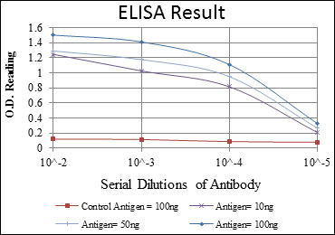
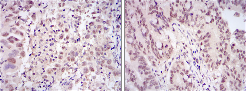
Applications |
WB, IHC, E
|
||
Dilution |
E~~1/10000
WB~~1/500 - 1/2000
IHC~~1/500 - 1/2000
|
||
Synonyms |
Cyclin-dependent kinase 9, 2.7.11.22, 2.7.11.23, C-2K, Cell division cycle 2-like protein kinase 4, Cell division protein kinase 9, Serine/threonine-protein kinase PITALRE, Tat-associated kinase complex catalytic subunit, CDK9, CDC2L4, TAK
|
||
Images |
|
||
Specification |
|||
Quantity |
|
||
| Select | Brand | Catalog No. | Product Name | Pack Size | Type | Field of Research | Specification | Quantity | Price(USD) | |
| 1 | Leading Biology | APG02467G | CCK4 / PTK7 Antibody (clone 4F9) | 50 μl | Monoclonal Antibodies |
|
$495.00 | Add Ask | ||
| 2 | Leading Biology | AMM04683G | GALT Antibody (clone 4C11) | 50 μg | Monoclonal Antibodies |
|
$545.00 | Add Ask | ||
| 3 | Leading Biology | AMM01402G | Vimentin (Mesenchymal Cell Marker) Antibody - With BSA and Azide | 50 ug | Monoclonal Antibodies |
|
$395.00 | Add Ask | ||
| 4 | Leading Biology | APR08280G | LTA4H / LTA4 Antibody (clone 9G8) | 50 μl | Monoclonal Antibodies |
|
$495.00 | Add Ask | ||
| 5 | Leading Biology | AMM00172G | CD1a / HTA1 (Mature Langerhans Cells Marker) Antibody - With BSA and Azide | 50 ug | Monoclonal Antibodies |
|
$395.00 | Add Ask | ||
| 6 | Leading Biology | AMM05750G | CEBPA Antibody | 100 μl | Monoclonal Antibodies |
|
$545.00 | Add Ask |

You have 0 item in your cart
You have 0 item in your inquiry list