> Antigen, Antibodies, ELISA, Western Blot > Primary Antibody > Monoclonal Antibodies > PDPK1 AntibodyBrand |
Leading Biology | Catalog Number |
AMM03042G |
Product Type |
Monoclonal Antibodies | Field of Research |
|
Product Overview |
We constantly strive to ensure we provide our customers with the best antibodies. As a result of this work we offer this antibody in purified format.
We are in the process of updating our datasheets. If you have any questions regarding this update, please feel free to contact our technical support team.
This product is a high quality PDPK1 Antibody.
|
||
Molecular Weight |
63.2kDa
|
||
Cellular Localization |
Antigen Cellular Localization:
Cytoplasm. Nucleus. Cell membrane; Peripheral membrane protein. Cell junction, focal adhesion Note=Tyrosine phosphorylation seems to occur only at the cell membrane. Translocates to the cell membrane following insulin stimulation by a mechanism that involves binding to GRB14 and INSR. SRC and HSP90 promote its localization to the cell membrane Its nuclear localization is dependent on its association with PTPN6 and its phosphorylation at Ser-396. Restricted to the nucleus in neuronal cells while in non-neuronal cells it is found in the cytoplasm. The Ser-241 phosphorylated form is distributed along the perinuclear region in neuronal cells while in non- neuronal cells it is found in both the nucleus and the cytoplasm IGF1 transiently increases phosphorylation at Ser-241 of neuronal PDPK1, resulting in its translocation to other cellular compartments. The tyrosine-phosphorylated form colocalizes with PTK2B in focal adhesions after angiotensin II stimulation
|
||
Host |
Mouse
|
||
Species Reactivity |
Human, Mouse, Rat, Monkey
|
||
Clone |
7E4G11
|
||
Isotype |
IgG1
|
||
Symbol |
PDK1
|
||
GeneID |
|||
UniProt ID |
|||
Function |
Serine/threonine kinase which acts as a master kinase, phosphorylating and activating a subgroup of the AGC family of protein kinases. Its targets include: protein kinase B (PKB/AKT1, PKB/AKT2, PKB/AKT3), p70 ribosomal protein S6 kinase (RPS6KB1), p90 ribosomal protein S6 kinase (RPS6KA1, RPS6KA2 and RPS6KA3), cyclic AMP-dependent protein kinase (PRKACA), protein kinase C (PRKCD and PRKCZ), serum and glucocorticoid-inducible kinase (SGK1, SGK2 and SGK3), p21-activated kinase-1 (PAK1), protein kinase PKN (PKN1 and PKN2). Plays a central role in the transduction of signals from insulin by providing the activating phosphorylation to PKB/AKT1, thus propagating the signal to downstream targets controlling cell proliferation and survival, as well as glucose and amino acid uptake and storage. Negatively regulates the TGF-beta-induced signaling by: modulating the association of SMAD3 and SMAD7 with TGF-beta receptor, phosphorylating SMAD2, SMAD3, SMAD4 and SMAD7, preventing the nuclear translocation of SMAD3 and SMAD4 and the translocation of SMAD7 from the nucleus to the cytoplasm in response to TGF-beta. Activates PPARG transcriptional activity and promotes adipocyte differentiation. Activates the NF-kappa-B pathway via phosphorylation of IKKB. The tyrosine phosphorylated form is crucial for the regulation of focal adhesions by angiotensin II. Controls proliferation, survival, and growth of developing pancreatic cells. Participates in the regulation of Ca(2+) entry and Ca(2+)-activated K(+) channels of mast cells. Essential for the motility of vascular endothelial cells (ECs) and is involved in the regulation of their chemotaxis. Plays a critical role in cardiac homeostasis by serving as a dual effector for cell survival and beta-adrenergic response. Plays an important role during thymocyte development by regulating the expression of key nutrient receptors on the surface of pre-T cells and mediating Notch-induced cell growth and proliferative responses. Provides negative feedback inhibition to toll-like receptor-mediated NF- kappa-B activation in macrophages. Isoform 3 is catalytically inactive.
|
||
Storage & Stability |
Store at +4°C short term. For long-term storage, aliquot and store at -20°C or below. Stable for 12 months at -20°C. Avoid repeated freeze-thaw cycles.
|
||
Applications |
WB, IHC, FC, ICC, E
|
||
Dilution |
E~~1/10000
WB~~1/500 - 1/2000
IF~~1/50 - 1/200
FC~~1/200 - 1/400
IHC~~1/200 - 1/1000
|
||
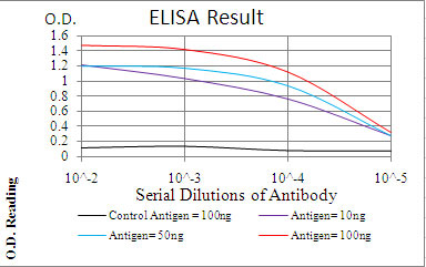
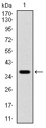
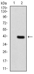
Images |
|
||
Specification |
|||
Quantity |
|
||
| Select | Brand | Catalog No. | Product Name | Pack Size | Type | Field of Research | Specification | Quantity | Price(USD) | |
| 1 | Leading Biology | APG02467G | CCK4 / PTK7 Antibody (clone 4F9) | 50 μl | Monoclonal Antibodies |
|
$495.00 | Add Ask | ||
| 2 | Leading Biology | AMM04683G | GALT Antibody (clone 4C11) | 50 μg | Monoclonal Antibodies |
|
$545.00 | Add Ask | ||
| 3 | Leading Biology | AMM01402G | Vimentin (Mesenchymal Cell Marker) Antibody - With BSA and Azide | 50 ug | Monoclonal Antibodies |
|
$395.00 | Add Ask | ||
| 4 | Leading Biology | APR08280G | LTA4H / LTA4 Antibody (clone 9G8) | 50 μl | Monoclonal Antibodies |
|
$495.00 | Add Ask | ||
| 5 | Leading Biology | AMM00172G | CD1a / HTA1 (Mature Langerhans Cells Marker) Antibody - With BSA and Azide | 50 ug | Monoclonal Antibodies |
|
$395.00 | Add Ask | ||
| 6 | Leading Biology | AMM05750G | CEBPA Antibody | 100 μl | Monoclonal Antibodies |
|
$545.00 | Add Ask |

You have 0 item in your cart
You have 0 item in your inquiry list