> Antigen, Antibodies, ELISA, Western Blot > Primary Antibody > Monoclonal Antibodies > NCOA3 AntibodyBrand |
Leading Biology | Catalog Number |
AMM02688G |
Product Type |
Monoclonal Antibodies | Field of Research |
|
Product Overview |
We constantly strive to ensure we provide our customers with the best antibodies. As a result of this work we offer this antibody in purified format.
We are in the process of updating our datasheets. If you have any questions regarding this update, please feel free to contact our technical support team.
This product is a high quality NCOA3 Antibody.
|
||
Molecular Weight |
155293 Da
|
||
Cellular Localization |
Antigen Cellular Localization:
Cytoplasm. Nucleus. Note=Mainly cytoplasmic and weakly nuclear. Upon TNF activation and subsequent phosphorylation, it translocates from the cytoplasm to the nucleus
|
||
Host |
Mouse
|
||
Species Reactivity |
Human
|
||
Clone |
2C11B12
|
||
Isotype |
IgG1
|
||
Symbol |
AIB1, BHLHE42, RAC3, TRAM1
|
||
GeneID |
|||
UniProt ID |
|||
Function |
Nuclear receptor coactivator that directly binds nuclear receptors and stimulates the transcriptional activities in a hormone-dependent fashion. Plays a central role in creating a multisubunit coactivator complex, which probably acts via remodeling of chromatin. Involved in the coactivation of different nuclear receptors, such as for steroids (GR and ER), retinoids (RARs and RXRs), thyroid hormone (TRs), vitamin D3 (VDR) and prostanoids (PPARs). Displays histone acetyltransferase activity. Also involved in the coactivation of the NF-kappa-B pathway via its interaction with the NFKB1 subunit.
|
||
Storage & Stability |
Store at +4°C short term. For long-term storage, aliquot and store at -20°C or below. Stable for 12 months at -20°C. Avoid repeated freeze-thaw cycles.
|
||
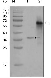
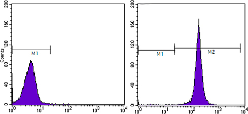
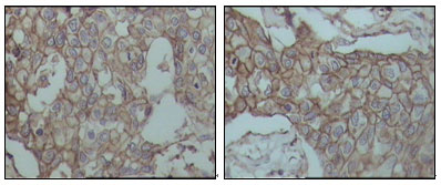
Applications |
WB, E
|
||
Dilution |
WB~~1/500 - 1/2000
FC~~1:200~~400
IHC~~1:200~~1000
|
||
Images |
|
||
Specification |
|||
Quantity |
|
||
| Select | Brand | Catalog No. | Product Name | Pack Size | Type | Field of Research | Specification | Quantity | Price(USD) | |
| 1 | Leading Biology | APG02467G | CCK4 / PTK7 Antibody (clone 4F9) | 50 μl | Monoclonal Antibodies |
|
$495.00 | Add Ask | ||
| 2 | Leading Biology | AMM04683G | GALT Antibody (clone 4C11) | 50 μg | Monoclonal Antibodies |
|
$545.00 | Add Ask | ||
| 3 | Leading Biology | AMM01402G | Vimentin (Mesenchymal Cell Marker) Antibody - With BSA and Azide | 50 ug | Monoclonal Antibodies |
|
$395.00 | Add Ask | ||
| 4 | Leading Biology | APR08280G | LTA4H / LTA4 Antibody (clone 9G8) | 50 μl | Monoclonal Antibodies |
|
$495.00 | Add Ask | ||
| 5 | Leading Biology | AMM00172G | CD1a / HTA1 (Mature Langerhans Cells Marker) Antibody - With BSA and Azide | 50 ug | Monoclonal Antibodies |
|
$395.00 | Add Ask | ||
| 6 | Leading Biology | AMM05750G | CEBPA Antibody | 100 μl | Monoclonal Antibodies |
|
$545.00 | Add Ask |

You have 0 item in your cart
You have 0 item in your inquiry list