> Antigen, Antibodies, ELISA, Western Blot > Primary Antibody > Monoclonal Antibodies > HLA-DRB (MHC II) Antibody - Without BSA and AzideBrand |
Leading Biology | Catalog Number |
AMM00846G |
Product Type |
Monoclonal Antibodies | Field of Research |
|
Product Overview |
We constantly strive to ensure we provide our customers with the best antibodies. As a result of this work we offer this antibody in purified format.
We are in the process of updating our datasheets. If you have any questions regarding this update, please feel free to contact our technical support team.
This product is a high quality HLA-DRB (MHC II) Antibody - Without BSA and Azide.
|
||
Molecular Weight |
~28kDa (beta chain)
|
||
Cellular Localization |
Antigen Cellular Localization:
Cell membrane; Single-pass type I membrane protein Endoplasmic reticulum membrane; Single-pass type I membrane protein Golgi apparatus, trans-Golgi network membrane; Single-pass type I membrane protein. Endosome membrane; Single-pass type I membrane protein. Lysosome membrane; Single-pass type I membrane protein. Late endosome membrane; Single-pass type I membrane protein. Note=The MHC class II complex transits through a number of intracellular compartments in the endocytic pathway until it reaches the cell membrane for antigen presentation
|
||
Host |
Mouse
|
||
Species Reactivity |
Human
|
||
Clone |
SPM289
|
||
Isotype |
Mouse / IgG2b, kappa
|
||
Symbol |
HLA-DRB2
|
||
GeneID |
|||
UniProt ID |
|||
Function |
Binds peptides derived from antigens that access the endocytic route of antigen presenting cells (APC) and presents them on the cell surface for recognition by the CD4 T-cells. The peptide binding cleft accommodates peptides of 10-30 residues. The peptides presented by MHC class II molecules are generated mostly by degradation of proteins that access the endocytic route, where they are processed by lysosomal proteases and other hydrolases. Exogenous antigens that have been endocytosed by the APC are thus readily available for presentation via MHC II molecules, and for this reason this antigen presentation pathway is usually referred to as exogenous. As membrane proteins on their way to degradation in lysosomes as part of their normal turn-over are also contained in the endosomal/lysosomal compartments, exogenous antigens must compete with those derived from endogenous components. Autophagy is also a source of endogenous peptides, autophagosomes constitutively fuse with MHC class II loading compartments. In addition to APCs, other cells of the gastrointestinal tract, such as epithelial cells, express MHC class II molecules and CD74 and act as APCs, which is an unusual trait of the GI tract. To produce a MHC class II molecule that presents an antigen, three MHC class II molecules (heterodimers of an alpha and a beta chain) associate with a CD74 trimer in the ER to form a heterononamer. Soon after the entry of this complex into the endosomal/lysosomal system where antigen processing occurs, CD74 undergoes a sequential degradation by various proteases, including CTSS and CTSL, leaving a small fragment termed CLIP (class-II-associated invariant chain peptide). The removal of CLIP is facilitated by HLA-DM via direct binding to the alpha-beta-CLIP complex so that CLIP is released. HLA-DM stabilizes MHC class II molecules until primary high affinity antigenic peptides are bound. The MHC II molecule bound to a peptide is then transported to the cell membrane surface. In B-cells, the interaction between HLA-DM and MHC class II molecules is regulated by HLA-DO. Primary dendritic cells (DCs) also to express HLA-DO. Lysosomal microenvironment has been implicated in the regulation of antigen loading into MHC II molecules, increased acidification produces increased proteolysis and efficient peptide loading.
|
||
Summary |
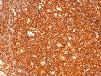
This MAb reacts with the beta-chain of HLA-DR antigen, a member of MHC class II molecules. It does not cross react with HLA-DP and HLA-DQ. The L243 antibody recognizes a different epitope than the SPM289 monoclonal antibody, and these antibodies do not cross-block binding to each other's respective epitopes. HLA-DR is a heterodimeric cell surface glycoprotein comprised of a 36kDa alpha (heavy) chain and a 28kDa beta (light) chain. It is expressed on B-cells, activated T-cells, monocytes/macrophages, dendritic cells and other non-professional APCs. In conjunction with the CD3/TCR complex and CD4 molecules, HLA-DR is critical for efficient peptide presentation to CD4+ T cells. It is an excellent histiocytic marker in paraffin sections producing intense staining. True histiocytic neoplasms are similarly positive. HLA-DR antigens also occur on a variety of epithelial cells and their corresponding neoplastic counterparts.
|
||
Form |
Liquid |
||
Storage & Stability |
Store at +4°C short term. For long-term storage, aliquot and store at -20°C or below. Stable for 12 months at -20°C. Avoid repeated freeze-thaw cycles.
|
||
Applications |
WB, IHC, IF, FC
|
||
Images |
|
||
Specification |
|||
Quantity |
|
||
| Select | Brand | Catalog No. | Product Name | Pack Size | Type | Field of Research | Specification | Quantity | Price(USD) | |
| 1 | Leading Biology | APG02467G | CCK4 / PTK7 Antibody (clone 4F9) | 50 μl | Monoclonal Antibodies |
|
$495.00 | Add Ask | ||
| 2 | Leading Biology | AMM04683G | GALT Antibody (clone 4C11) | 50 μg | Monoclonal Antibodies |
|
$545.00 | Add Ask | ||
| 3 | Leading Biology | AMM01402G | Vimentin (Mesenchymal Cell Marker) Antibody - With BSA and Azide | 50 ug | Monoclonal Antibodies |
|
$395.00 | Add Ask | ||
| 4 | Leading Biology | APR08280G | LTA4H / LTA4 Antibody (clone 9G8) | 50 μl | Monoclonal Antibodies |
|
$495.00 | Add Ask | ||
| 5 | Leading Biology | AMM00172G | CD1a / HTA1 (Mature Langerhans Cells Marker) Antibody - With BSA and Azide | 50 ug | Monoclonal Antibodies |
|
$395.00 | Add Ask | ||
| 6 | Leading Biology | AMM05750G | CEBPA Antibody | 100 μl | Monoclonal Antibodies |
|
$545.00 | Add Ask |

You have 0 item in your cart
You have 0 item in your inquiry list