> Antigen, Antibodies, ELISA, Western Blot > Primary Antibody > Monoclonal Antibodies > HINT1 AntibodyBrand |
Leading Biology | Catalog Number |
AMM02521G |
Product Type |
Monoclonal Antibodies | Field of Research |
|
Product Overview |
We constantly strive to ensure we provide our customers with the best antibodies. As a result of this work we offer this antibody in purified format.
We are in the process of updating our datasheets. If you have any questions regarding this update, please feel free to contact our technical support team.
This product is a high quality HINT1 Antibody.
|
||
Molecular Weight |
13802 Da
|
||
Cellular Localization |
Antigen Cellular Localization:
Cytoplasm. Nucleus. Note=Interaction with CDK7 leads to a more nuclear localization
|
||
Host |
Mouse
|
||
Species Reactivity |
Human, Mouse, Rat
|
||
Target |
This HINT1 antibody is generated from a mouse immunized with a recombinant protein of human HINT1.
|
||
Clone |
1500CT836.13.93
|
||
Isotype |
IgG1,k
|
||
Symbol |
HINT, PKCI1, PRKCNH1
|
||
GeneID |
|||
UniProt ID |
|||
Function |
Hydrolyzes purine nucleotide phosphoramidates with a single phosphate group, including adenosine 5'monophosphoramidate (AMP-NH2), adenosine 5'monophosphomorpholidate (AMP-morpholidate) and guanosine 5'monophosphomorpholidate (GMP-morpholidate). Hydrolyzes lysyl-AMP (AMP-N-epsilon-(N-alpha-acetyl lysine methyl ester)) generated by lysine tRNA ligase, as well as Met-AMP, His- AMP and Asp-AMP, lysyl-GMP (GMP-N-epsilon-(N-alpha-acetyl lysine methyl ester)) and AMP-N-alanine methyl ester. Can also convert adenosine 5'-O-phosphorothioate and guanosine 5'-O- phosphorothioate to the corresponding nucleoside 5'-O-phosphates with concomitant release of hydrogen sulfide. In addition, functions as scaffolding protein that modulates transcriptional activation by the LEF1/TCF1-CTNNB1 complex and by the complex formed with MITF and CTNNB1. Modulates p53/TP53 levels and p53/TP53-mediated apoptosis. Modulates proteasomal degradation of target proteins by the SCF (SKP2-CUL1-F-box protein) E3 ubiquitin- protein ligase complex.
|
||
Summary |
Hydrolyzes purine nucleotide phosphoramidates with a single phosphate group, including adenosine 5'monophosphoramidate (AMP-NH2), adenosine 5'monophosphomorpholidate (AMP-morpholidate) and guanosine 5'monophosphomorpholidate (GMP-morpholidate). Hydrolyzes lysyl-AMP (AMP-N-epsilon-(N-alpha-acetyl lysine methyl ester)) generated by lysine tRNA ligase, as well as Met-AMP, His- AMP and Asp-AMP, lysyl-GMP (GMP-N-epsilon-(N-alpha-acetyl lysine methyl ester)) and AMP-N-alanine methyl ester. Can also convert adenosine 5'-O-phosphorothioate and guanosine 5'-O- phosphorothioate to the corresponding nucleoside 5'-O-phosphates with concomitant release of hydrogen sulfide. In addition, functions as scaffolding protein that modulates transcriptional activation by the LEF1/TCF1-CTNNB1 complex and by the complex formed with MITF and CTNNB1. Modulates p53/TP53 levels and p53/TP53-mediated apoptosis. Modulates proteasomal degradation of target proteins by the SCF (SKP2-CUL1-F-box protein) E3 ubiquitin- protein ligase complex.
|
||
Storage & Stability |
Store at +4°C short term. For long-term storage, aliquot and store at -20°C or below. Stable for 12 months at -20°C. Avoid repeated freeze-thaw cycles.
|
||
Applications |
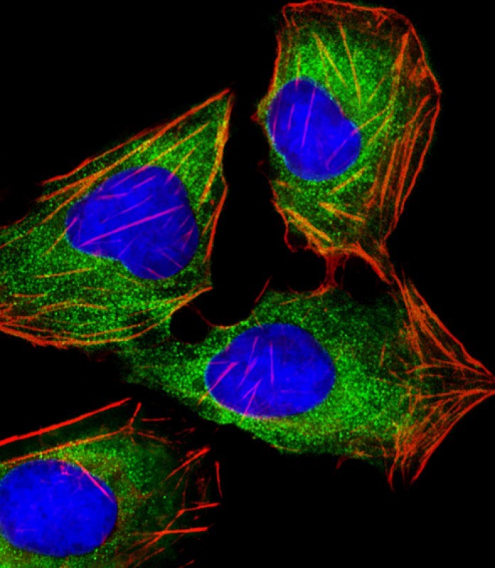
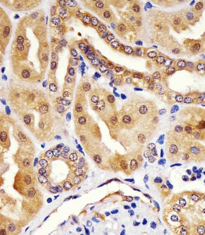
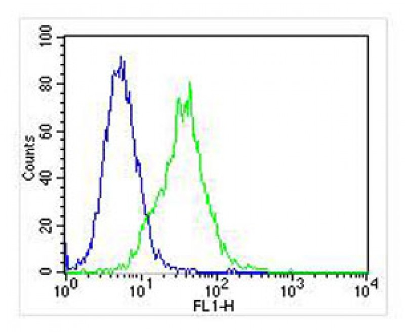
IF, IHC-P, FC, WB, E
|
||
Dilution |
IF~~1:25
IHC-P~~1:25
FC~~1:25
WB~~1:4000
|
||
Images |
|
||
Specification |
|||
Quantity |
|
||
| Select | Brand | Catalog No. | Product Name | Pack Size | Type | Field of Research | Specification | Quantity | Price(USD) | |
| 1 | Leading Biology | APG02467G | CCK4 / PTK7 Antibody (clone 4F9) | 50 μl | Monoclonal Antibodies |
|
$495.00 | Add Ask | ||
| 2 | Leading Biology | AMM04683G | GALT Antibody (clone 4C11) | 50 μg | Monoclonal Antibodies |
|
$545.00 | Add Ask | ||
| 3 | Leading Biology | AMM01402G | Vimentin (Mesenchymal Cell Marker) Antibody - With BSA and Azide | 50 ug | Monoclonal Antibodies |
|
$395.00 | Add Ask | ||
| 4 | Leading Biology | APR08280G | LTA4H / LTA4 Antibody (clone 9G8) | 50 μl | Monoclonal Antibodies |
|
$495.00 | Add Ask | ||
| 5 | Leading Biology | AMM00172G | CD1a / HTA1 (Mature Langerhans Cells Marker) Antibody - With BSA and Azide | 50 ug | Monoclonal Antibodies |
|
$395.00 | Add Ask | ||
| 6 | Leading Biology | AMM05750G | CEBPA Antibody | 100 μl | Monoclonal Antibodies |
|
$545.00 | Add Ask |

You have 0 item in your cart
You have 0 item in your inquiry list