> Antigen, Antibodies, ELISA, Western Blot > Primary Antibody > Monoclonal Antibodies > EPHB2 AntibodyBrand |
Leading Biology | Catalog Number |
AMM02490G |
Product Type |
Monoclonal Antibodies | Field of Research |
|
Product Overview |
We constantly strive to ensure we provide our customers with the best antibodies. As a result of this work we offer this antibody in purified format.
We are in the process of updating our datasheets. If you have any questions regarding this update, please feel free to contact our technical support team.
This product is a high quality EPHB2 Antibody.
|
||
Molecular Weight |
117493 Da
|
||
Cellular Localization |
Antigen Cellular Localization:
Cell membrane; Single-pass type I membrane protein. Cell projection, axon. Cell projection, dendrite
|
||
Host |
Mouse
|
||
Species Reactivity |
Human
|
||
Target |
Purified His-tagged EPHB2 protein(Fragment, between amino acids 127~425) was used to produce this monoclonal antibody.
|
||
Clone |
48CT12.6.4
|
||
Isotype |
IgG1κ
|
||
Symbol |
DRT, EPHT3, EPTH3, ERK, HEK5, TYRO5
|
||
GeneID |
|||
UniProt ID |
|||
Function |
Receptor tyrosine kinase which binds promiscuously transmembrane ephrin-B family ligands residing on adjacent cells, leading to contact-dependent bidirectional signaling into neighboring cells. The signaling pathway downstream of the receptor is referred to as forward signaling while the signaling pathway downstream of the ephrin ligand is referred to as reverse signaling. Functions in axon guidance during development. Involved in the guidance of commissural axons, that form a major interhemispheric connection between the 2 temporal lobes of the cerebral cortex. Also involved in guidance of contralateral inner ear efferent growth cones at the midline and of retinal ganglion cell axons to the optic disk. In addition to axon guidance, also regulates dendritic spines development and maturation and stimulates the formation of excitatory synapses. Upon activation by EFNB1, abolishes the ARHGEF15-mediated negative regulation on excitatory synapse formation. Controls other aspects of development including angiogenesis, palate development and in inner ear development through regulation of endolymph production. Forward and reverse signaling through the EFNB2/EPHB2 complex regulate movement and adhesion of cells that tubularize the urethra and septate the cloaca. May function as a tumor suppressor.
|
||
Summary |
Ephrin receptors and their ligands, the ephrins, mediate numerous developmental processes, particularly in the nervous system. Based on their structures and sequence relationships, ephrins are divided into the ephrin-A (EFNA) class, which are anchored to the membrane by a glycosylphosphatidylinositol linkage, and the ephrin-B (EFNB) class, which are transmembrane proteins. The Eph family of receptors are divided into 2 groups based on the similarity of their extracellular domain sequences and their affinities for binding ephrin-A and ephrin-B ligands. Ephrin receptors make up the largest subgroup of the receptor tyrosine kinase (RTK) family. The protein encoded by this gene is a receptor for ephrin-B family members.
|
||
Storage & Stability |
Store at +4°C short term. For long-term storage, aliquot and store at -20°C or below. Stable for 12 months at -20°C. Avoid repeated freeze-thaw cycles.
|
||
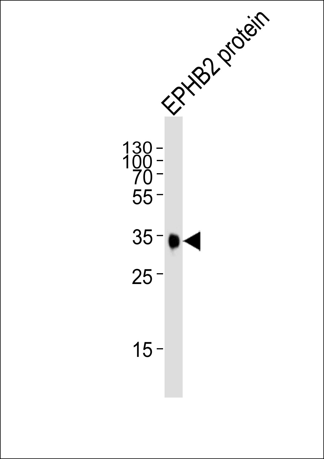
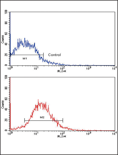
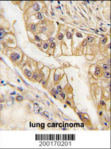
Applications |
FC, WB, IHC-P, E
|
||
Dilution |
WB~~1:1000
FC~~1:10~50
IHC-P~~1:200
|
||
Images |
|
||
Specification |
|||
Quantity |
|
||
| Select | Brand | Catalog No. | Product Name | Pack Size | Type | Field of Research | Specification | Quantity | Price(USD) | |
| 1 | Leading Biology | APG02467G | CCK4 / PTK7 Antibody (clone 4F9) | 50 μl | Monoclonal Antibodies |
|
$495.00 | Add Ask | ||
| 2 | Leading Biology | AMM04683G | GALT Antibody (clone 4C11) | 50 μg | Monoclonal Antibodies |
|
$545.00 | Add Ask | ||
| 3 | Leading Biology | AMM01402G | Vimentin (Mesenchymal Cell Marker) Antibody - With BSA and Azide | 50 ug | Monoclonal Antibodies |
|
$395.00 | Add Ask | ||
| 4 | Leading Biology | APR08280G | LTA4H / LTA4 Antibody (clone 9G8) | 50 μl | Monoclonal Antibodies |
|
$495.00 | Add Ask | ||
| 5 | Leading Biology | AMM00172G | CD1a / HTA1 (Mature Langerhans Cells Marker) Antibody - With BSA and Azide | 50 ug | Monoclonal Antibodies |
|
$395.00 | Add Ask | ||
| 6 | Leading Biology | AMM05750G | CEBPA Antibody | 100 μl | Monoclonal Antibodies |
|
$545.00 | Add Ask |

You have 0 item in your cart
You have 0 item in your inquiry list