Transmission Electron Microscopy
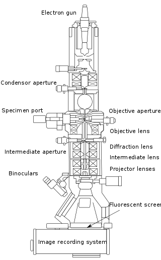
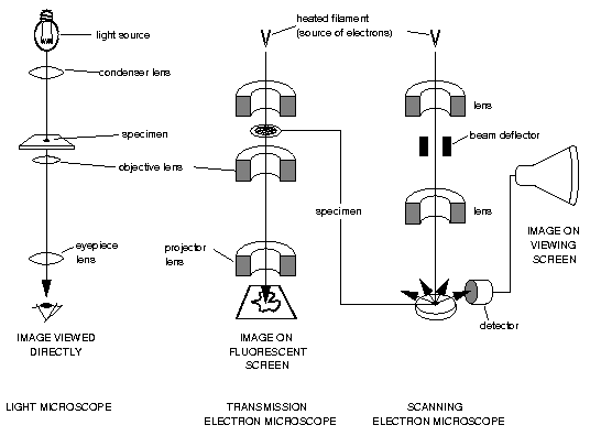
Transmission electron microscopy (TEM) is a microscopy technique in which a beam of electrons is transmitted through a specimen to form an image. The specimen is most often an ultrathin section less than 100 nm thick or a suspension on a grid. An image is formed from the interaction of the electrons with the sample as the beam is transmitted through the specimen. The image is then magnified and focused onto an imaging device, such as a fluorescent screen, a layer of photographic film, or a sensor such as a scintillator attached to a charge-coupled device. Transmission electron microscopes are capable of imaging at a significantly higher resolution than light microscopes, owing to the smaller de Broglie wavelength of electrons. This enables the instrument to capture fine detail—even as small as a single column of atoms, which is thousands of times smaller than a resolvable object seen in a light microscope. Transmission electron microscopy is a major analytical method in the physical, chemical and biological sciences.
TEM instruments boast an enormous array of operating modes including conventional imaging, diffraction, spectroscopy, and combinations of these. Even within conventional imaging, there are many fundamentally different ways that contrast is produced, called "image contrast mechanisms." Contrast can arise from position-to-position differences in the thickness or density, atomic number, crystal structure or orientation, the slight quantum-mechanical phase shifts that individual atoms produce in electrons that pass through them, the energy lost by electrons on passing through the sample and more. Each mechanism tells the user a different kind of information, depending not only on the contrast mechanism but on how the microscope is used—the settings of lenses, apertures, and detectors. For this reason, TEM is regarded as an essential tool for nanoscience in both biological and materials fields.

Introduction of Scanning Transmission Electron Microscopy
Scanning transmission electron microscopy (STEM) combines the principles of transmission electron microscopy (TEM) and scanning electron microscopy (SEM) and can be performed on either type of instrument, As with a transmission electron microscope (TEM), images are formed by electrons passing through a sufficiently thin specimen. A typical STEM is a conventional transmission electron microscope equipped with additional scanning coils, detectors, and necessary circuitry, which allows it to switch between operating as a STEM, or a CTEM; however, dedicated STEMs are also manufactured.
High resolution scanning transmission electron microscopes require exceptionally stable room environments. In order to obtain atomic-resolution images in STEM, the level of vibration, temperature fluctuations, electromagnetic waves, and acoustic waves must be limited in the room housing the microscope.

Advantages of TEM and STEM
Related Service
Why Leading Biology?
At Leading Biology, we custom protein purification design for every single protein to ensure the production and recovery rate as high as possible.
Working with us, you will get stability, and it means a reliable partner to help streamline your R&D process.
Working with us, you will get the guaranteed service to accommodate your requirements.
· Vigorous quality control system to ensure the required quality and reproducibility
· Competitive price with fast turnaround time
Contact Information
Please obtain a quote before ordering, and refer to the quote number when you place an order.
Orders are typically confirmed within 12 hours.
Have a Question? Email us info@leadingbiology.com
Order Products: Order Related Products
By Phone: 1-661-524(LBI)-0262 (USA)
| No | Headline | Click | Author | Date |
| 1 | siRNA/shRNA gene knockdown | 1737 | Leading Biology | 2018-01-26 |
| 2 | Tetracycline Induced Gene knockout/knockin | 1939 | Leading Biology | 2018-01-26 |
| 3 | New Generation Embryonic Stem Cells Gene Targeting | 1938 | Leading Biology | 2018-01-26 |
| 4 | TALEN Technology | 1781 | Leading Biology | 2018-01-26 |
| 5 | CRISPR-CAS Technology | 2204 | Leading Biology | 2018-01-26 |
| 6 | Microorganisms Gene Modification Services | 1673 | Leading Biology | 2018-01-26 |