> Information Center > Technical FAQs > Cell Culture Technology Column > Common contamination and solutions in cell culturesCommon contamination and solutions in cell cultures
Common types of biological pollution in cell culture include bacterial contamination, mold contamination, mycoplasma contamination, black dots contamination, fungal contamination, and protozoa contamination. The characteristics of these contamination in cell culture and the corresponding solutions are as follows:
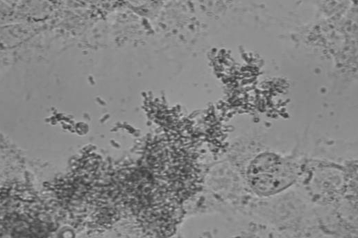
1. Bacteria contamination
The bacteria present black and fine sand under the ordinary inverted microscope. According to the different kinds of infected bacteria, it may show different shapes, and the culture solution will generally become cloudy and yellow, which has an obvious influence on cell growth.

Solutions: Check the sterilization of utensils carefully to ensure the ventilation time and the pressure of autoclaving is enough. Most of all, items such as pipettes that are in contact with culture medium may cause contamination of the storage solution if it is contaminated twice. So be sure to pay attention! Besides, check the culture medium turbidity before the next use. And, it is recommended to add antibiotics to the culture medium, such as tetracycline, gentamycin, or streptomycin.
2. Mold contamination
Normally, the culture medium is clear and transparent with no impurities under the inverted microscope even if it already has been cultured for two or three days under 37℃ in incubators. Once flocculent impurities occur, filamentous and conglobated floats are microscopically shown and the hyphae is obvious. At this time, although the bacterial cells are still growing, their vital state will gradually deteriorate after a long time.

Solutions: Wipe the CO2 incubator with copper sulfate solution, and add saturated amount of copper sulfate to the tray. Or adding the saturated disodium hydrogen phosphate high salinity solution after disinfection to avoid the mold contamination.
In case of the mold contamination, temporarily transfer all of cells, and immediately and thoroughly use peracetic acid solution to wipe the incubator including its baffle plate and inside wall. Place the peracetic acid in the incubator for an hour to diffuse the steam and transfer the bacteria cells to the incubator after the odor of peracetic acid is dissipated. It's worthwhile to note that the incubator needs regular cleaning every two months, especially in rainy seasons. Wiping incubators with 84 Disinfection Solution, clean water and 75 % alcohol successively and irradiate with ultraviolet light is also a useful method.
In order to prevent the mold contamination, it is necessary to add 3μg/ml amphotericin, mycostatin, actinomycin D or penicillin-streptomycin. Once contaminated, it’s impossible to recover, and it makes no difference to the matter even with aforementioned antibiotics. The best option for mycoplasma-infected cell cultures is to discard infected cultures and replace them with fresh stocks which are mycoplasma-free and disinfect the environment thoroughly. If all the bacteria cells are contaminated, it may be systemic contamination. Therefore, it is necessary to check the culture medium and experiment equipment. If it is only individual contamination, it may be an operational problem. So it is essential to pay attention to the specifications of the experimental operation steps.
3. Mycoplasma contamination
Mycoplasmas are covert contaminants, and it can't be removed by filtration. The absence of any visible signs of mycoplasma contamination, such as cytopathogenicity and changes in turbidity or pH (even in heavily contaminated cultures), leads to a false sense of security. The culture medium usually become turbid after mycoplasma contamination, and interiorly, serums are seldom tested for negative detection of mycoplasma, which is one of the most common microorganisms in bovine serum.

Solutions: Once contaminated by mycoplasma, especially for important cell lines, it is necessary to remove mycoplasma and general methods are antibiotic treatment, antiserum treatment and antibiotics treatment combined with antiserum and alexin. It is noteworthiness that the most prominent structural feature of mycoplasma is the absence of cell walls. Therefore, it is completely insensitive to biosynthetic antibiotics acting on the cell wall, such as β-lactams, vancomycin, etc., and is generally resistant to polymyxin, rifampicin, and sulfa drugs. Instead, tetracyclines and macrolides have the most effective antibiotics inhibitory against mycoplasma, while aminoglycosides and chloramphenicol have less inhibitory effects. Also, all cultures need to be replaced when necessary. Finally, the usual method of filtration sterilization has no effect on mycoplasma.
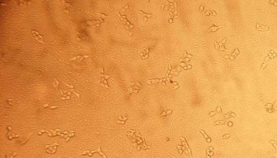
4. Black dots contamination
The black swimming dots can penetrate the filter membrane and spread through the air. They shows as black dota at low magnification and mobile black specks at high magnification. The culture solution is also not turbid, and generally affects less, so the bacteria cells can still be used. Usually, after black dots contamination, the cells grow well, and the moving objects are not evidently increased, and the color and transparency of the culture medium have no significant change. Such a similar phenomenon can be found in the same batch of serum-cultured cells.
Solutions: Black dots do not have a significant effect on cell growth status, and will naturally disappear after cell proliferation, so no special treatment is needed except for serum replacement. And it is suggested to increase the plate density for the higher survival rate of the cells if they are likely to be contaminated by the small moving dots.
5. Fungi contamination
After fungal contamination, the culture medium is generally clear and remains its original color. Filaments can be observed under the microscope. Initially, some fungi resemble dead cell debris, but many of them are clearly seen, and look like coraline, unlike cell debris indistinguishable. In addition, they will grow thin and black filament slowly because they grow more slower and are not as easily found as bacteria, once the medium are found to be contaminated, they will be difficult to survive.

Solutions: Once contaminated by fungi, discard it decisively, then sterilize the culture room, CO2 incubator, utensils and culture medium thoroughly.
6. Protozoa contamination
After the protozoa contamination, the cells culture medium becomes slightly turbid. Under the microscope, a large number of small dots move back and forth. Although the cells can still grow at this time, the reproduction speed slows down, and the cells have a bad growth state with unclear edges and become non-transparent. Protozoa and bacteria cells form a symbiotic relationship. Simultaneously, protozoa compete with cells for nutrition. This symbiosis is very common, but the cells predominate because the number of protozoa is relatively small, and thus having no effect on the normal growth of cells, only when they reach a certain amount, and eventually become malignant cycle.
Solution: There are many possible causes for protozoa contamination, such as liquid disinfection problems, operational problems, environmental problems, etc. If the cells are enough, discard the contaminated cell decisively and resuscitate cells. If preserved, purchase the relevant sterilization reagents.


