> Information Center > Technical FAQs > Autophagy Technology Column > How do immunohistochemistry and immunocytochemistry differImmunohistochemistry (IHC), Immunocytochemistry (ICC) and Immunofluorescence (IF) all utilize antibodies to provide visual details about protein abundance, distribution and localization. These terms are often confusing and are sometimes mistakenly used interchangeably. Thus, it is important to understand the fundamental differences between these various techniques.
cadherin antibody - Ecadherin antibody
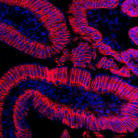
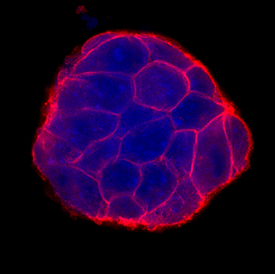
IHC-Fr (on left): Cadherin‑17 was detected in perfusion fixed frozen sections of mouse intestine using rabbit anti-mouse Cadherin‑17 monoclonal antibody, followed by the NorthernLights™ 557-conjugated anti-rabbit IgG secondary antibody and counterstained with DAPI (blue). Specific staining of Cadherin-17 was localized to plasma membranes. ICC/IF (on right): E‑Cadherin was detected in immersion fixed D3 mouse embryonic stem cell line using Goat Anti-Human/Mouse E‑Cadherin Antigen Affinity-purified Polyclonal Antibody at 10 µg/mL for 3 hours at room temperature. Cells were stained using the NorthernLights™ 557-conjugated Anti-Goat IgG Secondary Antibody and counterstained with DAPI (blue). Specific staining was localized to cell surfaces.
Sample Type - tissue vs cells –
As their names imply, IHC uses tissue sections, either paraffin embedded or frozen, whereas ICC refers to the staining of isolated or cultured intact cells. ICC samples may be from tissue culture cell lines (adherent or in suspension) as well as directly from a human or animal source. Prior to staining, IHC and ICC samples are also processed differently. A few examples of these differences include:
ICC samples, compared to IHC, undergo a shorter fixation period.
Fixatives, such as formaldehyde, can mask epitopes and reduce antigen-antibody binding. An antigen retrieval method is typically included before commencing IHC staining to restore tissue antigenicity.
In IHC, samples are either embedded in paraffin or frozen to preserve tissue morphology.
Labeling Method – chromogenic vs fluorescent – IHC and ICC have traditionally used chromogenic reagents to detect target antigens. However, with the extensive list of available fluorescent labels and their ease of use in multiplex applications, researchers more often use IF with ICC. Fluorescence detection for IHC is also gaining popularity.
In chromogenic detection, an enzyme such as horseradish peroxidase (HRP) converts a soluble substrate including 3,3'-Diaminobenzidine (DAB) and 3-amino-9-ethylcarbazole (AEC) into an insoluble colored product at the antigen site.
In IF detection, the fluorochrome conjugated reagent is bound, either directly or indirectly, to the primary antibody and, once excited, will emit light at a specific wavelength.
Note: Since IHC and ICC by themselves don’t specify the labeling method, it can help to combine them with the detection technique to clearly delineate the sample type and labeling method, i.e. ICC/IF or fluorescent IHC.
Looking for a validated secondary antibody to detect your antigen of interest? Novus Biologicals offers thousands of conjugated secondary antibodies including more than 40 different enzymatic, fluorescent, or biotin labels to choose from. Find the right detection reagent for each of your applications - Western blots, IHC, ICC, ELISAs.


